
Continuing from the previous part, we examine the remaining four pathways, from 4M Hardening to 7M Collapse, along with the integration of the 7M. The cited references follow the manuscript as written.
4. 4M Hardening: It Hardens
4M Hardening is the loss of elasticity through fibrosis and calcification. What ought to be pliable turns rigid. There are two types of arterial calcification. The distinction matters. The calcification that forms inside an atherosclerotic plaque is mainly a problem of the intima. The plaque calcification explained under 1M Obstruction & Rupture corresponds to this. The stiffening of the entire arterial wall in aging, diabetes, and kidney disease is centered on medial calcification. This is chiefly what 4M Hardening deals with. The two phenomena are different, but in that both are hardening made by calcium they wear the same face.
Let us look at arterial hardening in concrete terms. A healthy aorta is an elastic artery. Its media is rich in elastin, the elastic fiber. The elastic fibers grant the artery its elasticity. It can stretch and then shrink. When the heart contracts, blood is pushed out into the aorta. About 70 milliliters of blood pours out in 0.3 seconds. It is an enormous pressure. The elastic aorta expands. It absorbs the pressure. Systolic blood pressure is kept appropriate. When the heart relaxes, the expanded aorta returns to its original size. This recoil pushes blood toward the periphery. Blood flow is maintained even during diastole. This is the Windkessel effect. The aorta serves as an auxiliary pump. What happens when medial calcification progresses? The elastic fibers are replaced by calcification. Elastin is broken down and calcium is deposited in its place. The artery hardens. The aorta no longer expands. It cannot absorb the pressure. Systolic blood pressure rises. The aorta does not recoil. It cannot push the blood. Diastolic blood pressure falls relatively. The pulse pressure, that is, systolic blood pressure minus diastolic blood pressure, grows larger. For example, a blood pressure that was 120 over 80 becomes 160 over 70. The pulse pressure increases from 40 to 90.
An increase in pulse pressure brings on several consequences. A burden falls on the heart. It must push blood out against a high systolic pressure. The left ventricle hypertrophies. In the end heart failure develops. The end organs are damaged. The large pulse pressure is transmitted to the small blood vessels. The microvessels of the brain, kidney, and retina are damaged. Arterial stiffness is measured by pulse wave velocity. It is the speed at which the pulse wave propagates along the artery. In a stiffened artery the pulse wave propagates quickly. With aging, the carotid-femoral pulse wave velocity increases markedly. According to a study published in the Journal of Applied Physiology in 2008, the higher the pulse wave velocity, the more significantly the risk of cardiovascular events increases.
It is not calcification alone. Fibrosis too contributes to hardening. With aging, collagen accumulates. Collagen cross-linking increases. Advanced glycation end products accumulate in collagen. The collagen grows stiff. Elastin is broken down. Elastin is hardly regenerated. The elastin made at birth is used for a lifetime. With aging, elastin is broken down and not replaced. Fibrosis and calcification progress together. Calcium is deposited in fibrotic tissue. Calcification promotes fibrosis. It is a vicious cycle. The result is the same. It is hardening. Seen from an evolutionary standpoint, it is because of the essential nature of calcium. Calcium forms structure. Bone is hard because of calcium. A seashell is hard because of calcium. A coral reef is hard because of calcium. When calcium is deposited, a thing becomes hard. This is the essence of calcium. When calcium is deposited in tissue that ought to be pliable, the pliability disappears. An artery ought to be pliable. When calcium is deposited, it hardens. Hardening is the loss of adaptability. The environment changes. One must be pliable to respond. Once hardened, one cannot respond.
5. 5M Overflow & Burst: It Overflows and Grows Large
5M Overflow & Burst is the excessive proliferation or enlargement of cells. Calcium flows into the cell in excess and the cell proliferates or grows abnormally large. Tumors are included as well. Calcium is a key regulator of cell proliferation. For a cell to divide, it must go around the cell cycle. From the growth phase to the DNA replication phase, from the preparation phase to the division phase. At several stages of this process a calcium signal is needed. For a resting cell to enter the cell cycle, calcium is needed. What happens when a growth factor binds to its receptor? The receptor is activated. Calcium is released from the endoplasmic reticulum. Cytosolic calcium rises. The risen calcium binds to calmodulin. The calcium-calmodulin complex activates several enzymes. Transcription factors are activated. The genes needed for entry into the cell cycle are expressed. The cell enters the growth phase. Calcium is also needed when it moves from the growth phase to the DNA replication phase. DNA replication begins. Calcium is the switch of cell division. A review published in the Biochemical Society Transactions in 2012 laid out how the remodeling of calcium signaling is connected to several diseases.
Normally this switch is precisely regulated. It turns on only when needed. It turns off when not needed. But what happens when calcium homeostasis is disturbed? What if cytosolic calcium is chronically slightly elevated? The switch is always slightly on. The cell keeps receiving the signal to divide. The normal signals that suppress proliferation are ignored. The cell keeps dividing. When a cell grows large without dividing, it is called hypertrophy. Take cardiac hypertrophy as an example. In hypertension or valvular disease a burden is placed on the heart. Cardiac muscle cells do not divide. Adult cardiac muscle cells hardly divide at all. Instead they grow large. They hypertrophy. In cardiac hypertrophy the calcium signal plays a key role. When mechanical stress is placed on cardiac muscle cells, the calcium signal changes. In particular, a low-frequency, sustained elevation of calcium induces hypertrophy. Calcineurin is the key. Calcineurin is a calcium-dependent enzyme. A sustained elevation of calcium activates calcineurin. Calcineurin activates a transcription factor. The activated transcription factor enters the nucleus. Hypertrophy-related genes are expressed. The cardiac muscle cells grow large. At first it is a compensatory response. The heart grows large in order to bear the burden. But if it persists, it progresses to pathological hypertrophy. The cardiac muscle becomes fibrotic. Cardiac function declines. Heart failure develops.
Seen from an evolutionary standpoint, once again it is because calcium is a signaling molecule. Cell proliferation is essential to the maintenance of life. To grow, cells must divide. To repair damage, cells must divide. But it must be controlled. Uncontrolled proliferation is a tumor. Calcium is the switch of this process. When calcium rises, division begins. When calcium falls, division stops. What happens when this switch breaks down? What if it becomes stuck in the always-on state? The cell keeps dividing. It escapes control. It overflows. This is Overflow & Burst.
6. 6M Disconnection: It Is Cut and Dies
6M Disconnection is the severing of nerves, the occlusion of blood vessels, and the necrosis of tissue. What ought to be joined is cut. Cells die. Take synaptic loss as an example. The brain is the organ of connection. Its 86 billion neurons are connected by about 100 trillion synapses. These connections process information, store memory, and produce consciousness. In Alzheimer's disease the earliest change to occur is synaptic loss. Before nerve cells die, the synapses disappear first. Just as Selkoe, in a paper published in Science in 2002, defined Alzheimer's disease as a synaptic failure, the degree of synaptic loss correlates best with cognitive decline. In fact the Alzheimer's researcher Khachaturian has long proposed the calcium hypothesis as a core hypothesis to explain brain aging and Alzheimer's. His paper published in the Annals of the New York Academy of Sciences in 1989 put forward the view that the collapse of calcium homeostasis is itself the central axis leading to synaptic loss and nerve cell death.
How is calcium involved in synaptic loss? In Alzheimer's disease, amyloid beta accumulates near the synapse. This disturbs calcium homeostasis. Amyloid beta can make holes in the cell membrane. Calcium flows in through these holes. Or it can overactivate the NMDA receptor. The NMDA receptor is a channel that readily passes calcium. The result is a rise of postsynaptic calcium. The risen calcium damages the synaptic structure. Calpain is activated. Calpain is a calcium-dependent protease. Calpain breaks down the structural proteins of the synapse. Dendritic spines shrink. Synapses are lost. At the same time synaptic strength weakens. Functionally too the connection weakens. The connection is cut. Memory disappears.
When a blood vessel is completely blocked, the tissue it supplied dies. In a myocardial infarction, when a coronary artery is blocked, blood flow to the cardiac muscle is cut off. Within a few minutes the energy source, adenosine triphosphate, is depleted. The ion pumps cannot work. Sodium accumulates inside the cell. The sodium-calcium exchanger works in reverse. Calcium enters the cell. Intracellular calcium is overloaded. The mitochondria take up calcium. When mitochondrial calcium is overloaded, the mitochondrial permeability transition pore opens. A review published in the Journal of Molecular and Cellular Cardiology in 2015 laid out how this mitochondrial permeability transition is the crux of ischemia-reperfusion injury. Cell death signals are released. The cell dies. The tissue becomes necrotic. The connection is cut.
Seen from an evolutionary standpoint, calcium is a double-edged sword. At normal concentrations calcium sustains life. It makes nerve transmission possible. It makes muscle contraction possible. At excess concentrations calcium kills the cell. It activates proteases. It damages mitochondria. It induces cell death. As we saw in Chapter 1, this is why the cell keeps its calcium concentration ten thousand times lower. Calcium is dangerous. That is why it is useful. When a dangerous substance is used as a signal, the signal is clear. But because it is a dangerous substance, when the regulation breaks down it is fatal. When calcium flows into the cell in excess, the cell dies. The connection is cut. This is Disconnection.
7. 7M Collapse: It Falls Apart
7M Collapse is the falling apart of the structure of bone and teeth. What ought to hold up gives way. The 7M runs in a direction different from the other six. 1M through 6M occur because calcium piles up in the wrong place. 7M occurs because calcium leaves the place where it should be, the bone and teeth. Yet 7M and 1M through 6M are two sides of the same process. The calcium that leaves in 7M piles up in 1M through 6M. Osteoporosis is a disease in which bone density decreases and the microstructure is damaged, so that the risk of fracture rises. Bone is the storehouse of calcium. Ninety-nine percent of the body's calcium is in the bone. It is about 1 to 1.2 kilograms. This calcium maintains the strength of the bone. The DIAH triggers pull calcium out of the bone. Osteoclasts resorb the bone. Calcium is released into the blood. Osteoblasts form new bone but cannot keep up with the amount resorbed. A net loss occurs. Bone density decreases. The microstructure of the bone is damaged. The trabeculae of the spongy bone grow thin and break. The cortical bone grows thin. The bone weakens. Even a slight impact brings on a fracture.
The consequences of a hip fracture are severe. According to an epidemiological review published in The Lancet in 2002, the one-year mortality of hip fracture patients aged 65 and over is about 20 to 30 percent. Many of the survivors lose the ability to live independently. The risk of admission to a nursing home rises. Upright walking is a defining trait of the human being. When a hip fracture makes walking impossible, an essential human function is lost. Teeth too have calcium as their main component. Enamel is composed of hydroxyapatite crystals. Periodontitis, that is, gum disease, is a leading cause of tooth loss. In periodontitis the alveolar bone, the bone of the gums, is destroyed. The DIAH triggers, inflammation in particular, promote resorption of the alveolar bone. When the alveolar bone is lost, the structure that supported the tooth gives way. The tooth loosens. In the end it falls out. Osteoporosis and periodontitis are connected. In a person with low systemic bone density, alveolar bone loss is also severe. Both are the result of the DIAH-7M pathway.
Seen from an evolutionary standpoint, it is because calcium made structure. When what was made is drawn out, it falls apart. Bone is the storehouse of calcium. For a lifetime it stored calcium, gave it out when needed, and filled it up again. But in the end it gives out too much and its own self falls apart. The substance that was the medium of birth becomes the medium of death. The substance that made structure brings structure down. The cycle is completed.
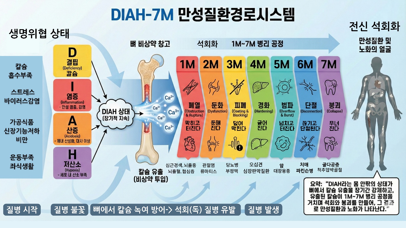
The Integration of the 7M: The Exit Mediated by Calcium
Until now we have looked at each of the 7M in turn. Now let us integrate them. 1M Obstruction & Rupture is calcium depositing in blood vessels so that the lumen is occluded, ruptures, and hemorrhage occurs. 2M Dysfunction is calcium depositing in joints and valves so that joint, muscle, and valve function declines. 3M Coating & Blocking is intracellular calcium rising so that receptors are blocked, signals are blocked, and insulin resistance occurs. 4M Hardening is calcium depositing in soft tissue so that fibrosis, calcification, and loss of elasticity occur. 5M Overflow & Burst is intracellular calcium becoming excessive so that cellular hyperproliferation, hypertrophy, and tumors occur. 6M Disconnection is intracellular calcium overloading so that nerves are severed, blood vessels are occluded, and necrosis occurs. 7M Collapse is calcium leaking out of the bone so that bone and tooth structure collapses. Seven different pathways. Seven different results. But they have a point in common. All of them are calcium. Calcium piles up, or calcium rises, or calcium leaves. The direction differs, but the substance is the same.
Why does evolution use one substance rather than many? It is because of efficiency. It is efficient to reuse a system that already exists. Calcium was already involved at every stage of life. In birth it activates the fertilized egg, in growth it forms the bone, in reproduction it transfers calcium to the fetus, and in signal transmission it regulates nerves, muscles, and hormones. There was no need to invent a new substance. When the whole cycle is controlled with a single substance, coordination is easy. When calcium absorption is reduced and efflux is increased, the seven pathways are activated at once. Without separate regulation the whole moves in the same direction.
1M through 6M and 7M are two sides of the same coin. The DIAH triggers pull calcium out of the bone. 7M begins. The calcium that has left piles up in the blood vessels. This is 1M. It piles up in the joints. This is 2M. It piles up in the organs. This is 4M. It enters into the cells. This is 3M, 5M, 6M. A deficiency on one side makes an excess on the other. The calcium that left the bone piles up in the soft tissue. This is the calcium paradox. In the bone, where it should be, there is none, and in the soft tissue, where it should not be, there it is. It is not a paradox. They are two aspects of the same process.
Conclusion: Calcium Is the Mediator of Exit
In this chapter we have looked at the 7M. But we have not simply listed pathological phenomena. We have argued. First, the calcium that has leaked out of the bone deposits in the soft tissue. This is not simply calcium passively piling up. It is an active cellular process. Vascular cells behave like osteoblasts. A genetic program is activated. Second, the calcium regulation of bone and soft tissue is connected. Regulators such as estrogen and vitamin K2 protect the bone while at the same time suppressing soft-tissue calcification. When this regulation weakens, both go the opposite way. This is the calcium paradox. Third, calcium carries out exit through seven pathways. It blocks the blood vessels, makes movement dull, blocks signals, removes elasticity, makes things escape control, cuts connections, and brings structure down. Fourth, at the center of every pathway there is calcium. A single substance mediates seven pathways. This is the efficiency of evolution. It reuses a system that already exists. The important point here is that this system is not a simplistic causal theory claiming that "calcium is the single cause of every disease." Rather, it is an interpretive frame that lays out how countless risk factors, such as stress, dietary habits, genetics, and environmental toxins, converge at the final stage upon one common pathway, namely DIAH, and upon the calcium that has left the bone and the 7M mechanisms.
Why calcium of all things? Calcium has been with life from its very beginning. What wakes the fertilized egg is a calcium wave. Calcium takes charge of both signal and structure. It transmits intracellular signals and it forms bone. Calcium is dangerous. That is why it is clear as a signal. And when it becomes excessive it is fatal. Calcium circulates. It is stored in the bone, then goes to the blood, to the soft tissue, and after death back to the environment. Is there any substance other than calcium that has all these properties? There is none. That is why calcium is the medium of evolution. From birth to death. It is the substance that runs through the whole of the cycle.
References
1. Shanahan, C. M., et al. (2011). Arterial calcification in chronic kidney disease: key roles for calcium and phosphate. Circulation Research, 109(6), 697-711. doi:10.1161/CIRCRESAHA.110.220509
2. Schurgers, L. J., et al. (2013). Vitamin K-dependent carboxylation of matrix Gla-protein: a crucial switch to control ectopic mineralization. Trends in Molecular Medicine, 19(4), 217-226. doi:10.1016/j.molmed.2013.01.001
3. Demer, L. L., & Tintut, Y. (2008). Vascular calcification: pathobiology of a multifaceted disease. Circulation, 117(22), 2938-2948. doi:10.1161/CIRCULATIONAHA.107.743161
4. Rosenthal, A. K., & Ryan, L. M. (2016). Calcium pyrophosphate deposition disease. The New England Journal of Medicine, 374(26), 2575-2584. doi:10.1056/NEJMra1511117
5. Dweck, M. R., et al. (2012). Calcific aortic stenosis: a disease of the valve and the myocardium. Journal of the American College of Cardiology, 60(19), 1854-1863. doi:10.1016/j.jacc.2012.02.093
6. Draznin, B. (2006). Molecular mechanisms of insulin resistance: serine phosphorylation of insulin receptor substrate-1 and increased expression of p85α. Diabetes, 55(8), 2392-2397. doi:10.2337/db06-0391
7. Mitchell, G. F. (2008). Effects of central arterial aging on the structure and function of the peripheral vasculature. Journal of Applied Physiology, 105(5), 1652-1660. doi:10.1152/japplphysiol.90549.2008
8. Berridge, M. J. (2012). Calcium signalling remodelling and disease. Biochemical Society Transactions, 40(2), 297-309. doi:10.1042/BST20110766
9. Halestrap, A. P., & Richardson, A. P. (2015). The mitochondrial permeability transition: a current perspective on its identity and role in ischaemia/reperfusion injury. Journal of Molecular and Cellular Cardiology, 78, 129-141. doi:10.1016/j.yjmcc.2014.08.018
10. Selkoe, D. J. (2002). Alzheimer's disease is a synaptic failure. Science, 298(5594), 789-791. doi:10.1126/science.1074069
11. Khachaturian, Z. S. (1989). Calcium hypothesis of Alzheimer's disease and brain aging. Annals of the New York Academy of Sciences, 568(1), 1-11. doi:10.1111/j.1749-6632.1989.tb12485.x
12. Cummings, S. R., & Melton, L. J. (2002). Epidemiology and outcomes of osteoporotic fractures. The Lancet, 359(9319), 1761-1767. doi:10.1016/S0140-6736(02)08657-9
13. Williams, G. C. (1957). Pleiotropy, natural selection, and the evolution of senescence. Evolution, 11(4), 398-411. doi:10.1111/j.1558-5646.1957.tb02911.x
14. Kirkwood, T. B. (1977). Evolution of ageing. Nature, 270(5635), 301-304. doi:10.1038/270301a0
15. Greenland, P., et al. (2018). Coronary calcium score and cardiovascular risk. Journal of the American College of Cardiology, 72(4), 434-447. doi:10.1016/j.jacc.2018.05.027