
This piece is the complete Chapter 1 of Calcium: The Reality of Evolution That Darwin and the Genome Map Missed (Yoon Jong-won). The body, figures, and citations follow the manuscript as written, and the subheadings are the subheadings found within the original.
Introduction: The Question a Single Number Poses
Anyone who has studied the life sciences has probably memorized this number at least once. The concentration of free calcium in the cytoplasm, about 100 nanomolar. We simply memorize this number that appears in the textbook and move on. Because it shows up on the exam. But let us pause for a moment and think hard about what this number means. Why exactly 100 nanomolar? Why not 1 micromolar, and not 10 nanomolar either, but precisely this range? And here an even more astonishing question arises. From Escherichia coli to the human neuron, why do all living things that branched apart over an unimaginable span of 3.8 billion years insist on this number? Bacteria and humans diverged from a common ancestor billions of years ago. In that time genes have changed countless times, forms have become completely different, and habitats have split to opposite extremes. There are archaea that live beside deep-sea hydrothermal vents spewing water close to 400 degrees, there are yaks that live breathing thin oxygen at a Himalayan altitude of 8,000 meters, and there are fish that swim in below-freezing seawater beneath the Antarctic ice sheet. The cells of these organisms operate under completely different conditions. And yet the cytoplasmic calcium concentration alone is almost identical. This is by no means a coincidence. Hidden here is the most fundamental secret of life, and understanding that secret becomes the first step toward understanding the relationship between calcium and evolution that runs through this entire book.
The Extreme Difference Between the Inside and the Outside of the Cell
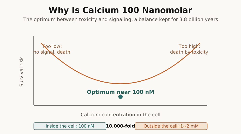
If we express in another way the fact that the cytoplasmic calcium concentration is about 100 nanomolar, we can grasp just how low a concentration this is. 100 nanomolar corresponds to 0.0001 millimolar, which means that in 1 liter of water only about 4 micrograms of calcium ions are dissolved. Of course, depending on the type of cell there is some variation between 50 nanomolar and 200 nanomolar, but the fact that almost all cells on Earth converge within this narrow range is something that many cell biology research institutions have confirmed repeatedly over several decades. Now, outside the cell, that is, in the extracellular fluid, the calcium concentration is about 1 millimolar to 2 millimolar. The calcium concentration in human blood corresponds roughly to this range, and the calcium concentration of seawater is also about 10 millimolar, far higher than inside the cell. When we place these two numbers side by side and compare them, an astonishing fact is revealed. The difference in calcium concentration between the inside and the outside of the cell reaches ten thousand-fold to twenty thousand-fold. What does it mean that, across a thin lipid bilayer only about 7 nanometers thick called the cell membrane, the calcium concentration differs by fully more than ten thousand-fold? This is like an enormous dam hundreds of meters high with the water levels on its two sides spread apart to an extreme. Water naturally tends to flow from a high place to a low place, and that force becomes more powerful the greater the difference in water level. In the same way, calcium ions too are constantly pressing to flow from outside the cell, where the concentration is high, into the cell, where the concentration is low, and this pressure is more powerful than we can imagine. To pump calcium out of the cell against this enormous concentration gradient is by no means an easy task, and for the sake of survival the cell must pour a vast amount of energy into this work.
To maintain this extreme concentration difference, the cell keeps several kinds of elaborate molecular machines running without rest. The calcium pump embedded in the cell membrane, the protein called PMCA, consumes one molecule at a time of the energy currency called ATP and pushes calcium ions out of the cell one by one. To send out a single calcium ion this pump must hydrolyze one molecule of ATP, and since one molecule of ATP stores about 30.5 kilojoules per mole of energy, this is by no means a cheap task. The SERCA pump on the membrane of the endoplasmic reticulum sequesters cytoplasmic calcium into the endoplasmic reticulum, a calcium storehouse inside the cell, and this pump too consumes ATP, using one molecule of ATP each time it puts two calcium ions into the endoplasmic reticulum. The mitochondria, the power plants of the cell, also absorb calcium and play a buffering role, and the sodium-calcium exchanger operates by taking three sodium ions into the cell in exchange for sending one calcium ion out of the cell, a system that plays a decisive role in regulating the rhythm of the heartbeat, especially in cardiac cells. All of these systems run without resting for a single moment, 24 hours a day, 365 days a year. Even while we sleep, and even in the cells of a hibernating animal, these pumps do not stop. According to several biochemistry research institutions, the cell spends about 5 percent to 10 percent of its total energy solely on maintaining the calcium concentration, which is an enormous investment. Considering that the human brain consumes about 20 percent of total energy, we can guess how vast the energy that goes into maintaining the calcium concentration is. Why does the cell try to exclude calcium even at such an expensive price?
Before answering this question, if we compare it with other ions, the peculiarity of calcium is revealed even more sharply. In the case of sodium, the concentration outside the cell is about 140 millimolar and the concentration inside the cell is about 10 millimolar, so the concentration difference is about 14-fold. Maintaining this concentration difference also takes energy, but even so it is 14-fold. In the case of potassium, the concentration outside the cell is about 4 millimolar and the concentration inside the cell is about 140 millimolar, so this ion is conversely higher inside the cell, but even so the concentration difference is only about 35-fold. Calcium, however, is ten thousand-fold to twenty thousand-fold. What does the fact that calcium alone maintains more than ten thousand-fold, while the other major cations maintain concentration differences on the order of tens of times, tell us? It tells us that calcium control is, for the cell, a top-priority management item incomparable to the other ions. The concentration difference of sodium and potassium is important for nerve signal transmission and the regulation of cell volume, but the concentration difference of calcium is related to something far more fundamental than that. Why does the cell exclude calcium alone so thoroughly? The answer lies in the toxicity of calcium.
The Toxicity of Calcium: Why the Cell Fears Calcium
The cytoplasm is packed densely with molecules essential to the activities of life. ATP, ADP, DNA, RNA, and thousands of kinds of proteins. Among these, ATP, DNA, and RNA have one thing in common. They all contain phosphate groups. ATP is called adenosine triphosphate because three phosphate groups are linked like a chain to the nucleic acid base called adenosine, and when the bond between these phosphate groups is broken, energy is released and supplies fuel to all the activities of the cell. The backbone of DNA and RNA is also made of a structure in which phosphate groups and sugars are alternately linked, so these molecules that carry genetic information cannot exist without phosphate groups either. The problem is that when calcium meets a phosphate group, it forms a precipitate that does not dissolve in water. The solubility of calcium phosphate is extremely low, so when calcium ions and phosphate ions meet, a solid precipitate forms almost instantly. The hydroxyapatite that makes up our bones and teeth is precisely one kind of calcium phosphate, and the reason bone is so hard is precisely this insolubility of calcium phosphate. When calcium increases in the cytoplasm, the phosphate groups of ATP and DNA begin to bind with calcium and precipitate. A catastrophe like bone forming inside the cell takes place.
Here a question may arise. If ATP exists abundantly inside the cell at the millimolar level, would it not precipitate upon meeting calcium that is only 100 nanomolar? Fortunately, magnesium exists abundantly inside the cell. The cytoplasmic magnesium concentration is about 0.5 millimolar to 1 millimolar, thousands of times to ten thousand times higher than calcium. ATP is for the most part stabilized in the form of magnesium-ATP, bound to magnesium, so it does not easily react with calcium at low concentrations. Magnesium plays the role of a shield that protects ATP. But when the calcium concentration explodes, the situation changes completely. Because calcium has a higher affinity for phosphate groups than magnesium, when the calcium concentration rises high enough, it pushes magnesium aside and begins to bind with ATP. And once calcium binds with ATP, an insoluble precipitate forms and ATP can no longer be used.
Why is this fatal? ATP is the energy currency of the cell. Every activity of life consumes ATP. For a muscle to contract, ATP is needed in the process of releasing and rebinding the bond between actin and myosin; to synthesize a protein, energy equivalent to four molecules of ATP is needed each time one amino acid is linked; for a nerve to send a signal, ATP is needed to run the ion pumps of the neuronal membrane. Even the calcium pump that pumps calcium out of the cell uses ATP as fuel, so when ATP is depleted the calcium pump too stops, and when the calcium pump stops, more calcium flows in and precipitates more ATP, and a vicious cycle begins. And when calcium binds with the phosphate groups of ATP and precipitates, ATP can no longer be used as an energy source. The energy system of the cell is paralyzed. It is the same with DNA and RNA. When the phosphate backbone of these molecules that hold genetic information binds with calcium and its structure is distorted or precipitated, gene expression itself becomes impossible. When RNA polymerase cannot read DNA and ribosomes cannot translate mRNA, the cell can no longer make new proteins. An important study published in 2019 revealed this process in detail at the molecular level. According to several mitochondrial research institutions, when calcium phosphate granules form inside the mitochondria, Complex I of the electron transport chain is directly inhibited and the rate of ATP synthesis decreases markedly. Calcium overload directly attacks the mitochondria, the power plants of the cell.
The toxicity of calcium does not stop at phosphate precipitation. On the surface of proteins, negatively charged amino acids are strategically distributed, aspartic acid and glutamic acid being representative. The side chains of these amino acids have carboxyl groups, so they carry a negative charge at physiological pH, and this negative charge attracts positively charged calcium ions. Under normal circumstances, calcium ions bind temporarily to this site and then come off, changing the structure of the protein, and this acts as a signal. But when the calcium concentration rises abnormally, the situation changes completely. When calcium binds excessively to the surface of proteins, a single calcium ion binds simultaneously to two or more proteins and comes to play the role of a bridge, and because of this, different proteins begin to clump together with calcium as the medium. Protein aggregation occurs. Aggregated proteins lose their normal three-dimensional structure and lose their function. Enzymes can no longer catalyze chemical reactions, structural proteins cannot maintain the shape of the cell, and receptor proteins cannot detect signals. In severe cases the aggregates physically occupy space inside the cell, obstruct the movement of other molecules, and paralyze the functions of cellular organelles such as the endoplasmic reticulum and the Golgi apparatus. The amyloid plaques observed in Alzheimer's disease and the Lewy bodies observed in Parkinson's disease are precisely the products of such protein aggregation, and it is no coincidence that research showing calcium overload to be related to these neurodegenerative diseases has accumulated over recent decades.
The cell membrane too becomes a target of calcium overload's attack. The cell membrane is made of a phospholipid bilayer, and the head portion of the phospholipid molecule contains a phosphate group, so it carries a negative charge. At a normal calcium concentration, calcium ions bind appropriately to the phospholipid heads and actually contribute to the stability of the membrane. Calcium builds bridges between the phospholipids and makes the membrane firmer. But when the calcium concentration rises excessively, calcium binds excessively to the phospholipids and severely reduces the fluidity of the membrane. The cell membrane is originally fluid like a liquid, so proteins move freely within the membrane and vesicles can fuse with and separate from the membrane, but when the membrane becomes stiff, all of these processes are obstructed. In severe cases holes are punched in the membrane or the membrane itself physically ruptures. When the cell membrane is destroyed, the cell dies. The contents of the cell pour out, and the cell contents released in this way send danger signals to the surrounding cells and trigger an inflammatory response. This is not a simple accidental death. Calcium overload also pulls the trigger of apoptosis, that is, the signal that the time has now come for the cell to die, programmed cell death. When calcium accumulates excessively in the mitochondria, a hole opens in the mitochondrial membrane and a protein called cytochrome c is released, and this triggers a chain reaction of enzymes called caspases, and the program by which the cell breaks itself down goes into operation. Calcium is the signal of life and at the same time the signal of death. This duality is the essence of calcium, and it is this very duality that explains the unique position calcium occupies in life.
The place where calcium toxicity appears most dramatically is the brain. When a stroke occurs, a blood vessel is blocked or bursts and the blood supply to some region of the brain is cut off. Neurons deprived of oxygen and glucose cannot make ATP in their mitochondria, and without ATP the calcium pumps of the cell membrane stop working. When the pumps stop, calcium slowly begins to seep into the cell. At the same time, from the damaged cells suffering from a lack of energy, a neurotransmitter called glutamate is released excessively. Under normal circumstances glutamate is an excitatory neurotransmitter that plays the role of activating neurons, but when released excessively it brings on catastrophe. Glutamate activates the NMDA receptors of adjacent neurons, and the NMDA receptor is an ion channel that opens when glutamate binds and is at the same time a calcium channel. Through the activated NMDA receptors, calcium flows in in large quantities. But these cells too already lack ATP and cannot pump out the calcium that has flowed in. The calcium overload that begins this way sets off a chain reaction. The mitochondria absorb calcium and then are damaged by the overload, reactive oxygen species are generated in the damaged mitochondria and attack the molecules inside the cell, and in the end the cell dies. From the dead cell glutamate is released again and attacks the surrounding cells. Several neuroscience research institutions call this phenomenon excitotoxicity, and point to it as the main cause of the mass death of neurons over the hours to days following a stroke. The reason time is important in the treatment of stroke is precisely that this excitotoxic chain reaction must be blocked.
Resolving the Paradox: Poison Becomes Signal
Here a paradox appears. If calcium is this dangerous, why does the cell not exclude calcium completely? Would it not be all right to remove all the calcium channels from the cell membrane? Evolution has tested countless variations over the unimaginable span of billions of years, so why did not a single living thing that completely removed its calcium channels survive? There is a reason it could not be done. Paradoxically, because calcium is dangerous, it could become a perfect signaling molecule. Because it was a poison, it could become a medicine, and because it was a danger, it could become an opportunity. This is the wisdom that life discovered, and understanding this paradox is the key to understanding the essence of calcium.
What conditions must a good signaling system satisfy? Let us take a radio broadcast as an example. First, when there is no broadcast, it must be completely quiet. There must be no background noise. If a crackling sound keeps being heard, it is hard to know whether an actual broadcast has begun. Second, when a broadcast begins, it must be clearly detected. If the announcer's voice is buried in background noise and cannot be heard, the broadcast is meaningless. Third, when a broadcast ends, it must quickly become quiet again. If the content of the previous broadcast keeps echoing, the next broadcast cannot be heard. In communications engineering this is called the signal-to-noise ratio, and the greater the strength of the signal relative to the strength of the noise, the better the quality of communication. Calcium perfectly satisfies all of these conditions. And the reason calcium can satisfy these conditions is precisely the toxicity of calcium.
What does it mean that the cytoplasmic calcium concentration is maintained at 100 nanomolar? If the concentration outside the cell is 1 millimolar to 2 millimolar, it means the concentration difference is ten thousand-fold to twenty thousand-fold. This is an enormous driving force. Just as, when you open the floodgate of a dam hundreds of meters high, the water pours down with tremendous force, the moment a calcium channel in the cell membrane opens, calcium is pushed sharply into the cell. When a single channel opens, the local concentration around it rises rapidly, and when the channel closes, the calcium pump immediately pumps the calcium out and the concentration falls again. According to several synapse research institutions, when a calcium channel opens at a synaptic terminal, in the space right beside the channel, within a radius of tens of nanometers, the calcium concentration soars in an instant from 10 micromolar to 100 micromolar. The difference between the background noise of 100 nanomolar and the signal of tens of micromolar reaches 100-fold to 1,000-fold. This means that the signal-to-noise ratio is extremely high. There is no chance at all of being confused about whether a signal has come. What would it have been like if the cytoplasmic calcium concentration were 1 micromolar? Then even if a signal came and the concentration became 10 micromolar, the difference would be only 10-fold. Because biological systems always have noise, with a 10-fold difference it could be hard to distinguish signal from noise. This is exactly why the cell excludes calcium at such an expensive price. Because calcium is a poison, it had to be excluded, and because it was thoroughly excluded, a perfect signaling system became possible.
The chemical properties of calcium are also ideal for signal transmission. The ionic radius of the calcium ion is about 1.00 angstrom, and this size fits exactly into the calcium-binding sites of proteins. Looking at the other elements that belong to the same Group 2 of the periodic table as calcium, magnesium has an ionic radius of 0.72 angstrom, too small; strontium is 1.18 angstrom, a bit large; and barium is 1.35 angstrom, too large. The calcium-binding sites of proteins have been optimized for calcium through hundreds of millions of years of evolution, so other ions cannot bind properly, or even if they bind they cannot induce the correct structural change. More important is the speed of binding. According to several biochemistry research institutions, calcium binds to proteins about 100 times faster than magnesium. This is related to the property of the hydration shell of calcium, that is, the layer of water molecules surrounding the calcium ion. Magnesium has a small ionic radius, so its charge density is high and it holds water molecules strongly, which makes it hard to shed the hydration shell.
Calcium, on the other hand, has a larger ionic radius, so its charge density is low and it can shed the hydration shell more easily. It binds quickly and comes off quickly. Thanks to this fast binding and dissociation speed, signal transmission on the order of milliseconds is possible. In a neuron, the time it takes for synaptic transmission to occur is about 1 millisecond. The electrical signal reaches the axon terminal, the calcium channel opens, calcium flows in, it binds to calmodulin, the synaptic vesicle fuses with the cell membrane, and the neurotransmitter is released: all of these processes must happen within 1 millisecond. Were it not for the fast binding speed of calcium, this would be impossible.
Calcium is also highly versatile. The coordination number of calcium is between 6 and 9, so it can bind simultaneously to 6 to 9 oxygen atoms. Thanks to this flexible mode of binding, calcium can bind to more than 200 kinds of different proteins. Magnesium has a fixed coordination number of 6, so the kinds of proteins it can bind to are limited. The most representative of the calcium-binding proteins is calmodulin. This small protein is made of 148 amino acids and has a dumbbell-shaped structure. Calmodulin has four calcium-binding sites, so it can bind to up to four calcium ions, and when calcium binds, the structure of calmodulin changes dramatically. Just as one closes a fist after opening a hand, when calmodulin binds with calcium it exposes a hydrophobic surface, and this surface binds with other proteins. Calmodulin whose structure has changed can activate more than 100 kinds of other proteins. Myosin light-chain kinase, which brings about muscle contraction; calcium-calmodulin-dependent kinase II, which is involved in the synaptic plasticity of neurons; adenylate cyclase, which makes cyclic AMP, the second messenger within the cell; nitric oxide synthase, which dilates blood vessels. Almost every function of the cell passes by way of calmodulin. And it is precisely calcium that turns on the switch of calmodulin. Calcium is the master switch of the vast factory that is the cell.
The Conservation of 3.8 Billion Years: The Importance Evolution Proves
Let us look at how universal the number of about 100 nanomolar for the cytoplasmic calcium concentration is. The cytoplasmic free calcium concentration of Escherichia coli is about 100 nanomolar. E. coli is a prokaryote, a simply structured cell with no nuclear membrane and no mitochondria. When the first cell appeared about 3.8 billion years ago, that cell probably looked similar to E. coli, and E. coli is one of the organisms that best preserves the form of that time. It is the same with yeast. Yeast is a eukaryote, so it has a nuclear membrane and mitochondria, but it is a single-celled organism that branched off from the ancestor of multicellular life about 1 billion years ago. The cytoplasmic calcium concentration of yeast is also about 100 nanomolar to 200 nanomolar. It is the same whether you look at plant cells, insect cells, fish cells, or mammalian cells.
None of them stray outside this narrow range. The human neuron, muscle cell, liver cell, and immune cell have completely different functions. The neuron transmits electrical signals, the muscle cell contracts to produce force, the liver cell is in charge of detoxification and metabolism, and the immune cell fights pathogens. But the baseline calcium concentration of all these cells is the same, about 100 nanomolar. The archaeon living beside water close to 400 degrees in the extreme environment of a deep-sea hydrothermal vent, the fish swimming in seawater at minus 2 degrees beneath the Antarctic ice sheet, the beetle enduring the hot sand of the Sahara Desert, and the hummingbird flying at the top of the Amazon rainforest all share the same number.
From the perspective of evolutionary biology, what such universal conservation means is profound. If a trait has been maintained almost unchanged for 3.8 billion years, it means that a very powerful selection pressure has existed. Selection pressure refers to the force by which the frequency of a particular trait changes over successive generations because individuals with that trait are more advantaged or disadvantaged in survival and reproduction than those without it. Individuals that could not maintain the cytoplasmic calcium concentration near 100 nanomolar were disadvantaged in survival and reproduction, and were weeded out over successive generations. That is why all the living things we observe today share this trait. There are not many traits that have been conserved this long in the history of life. The genetic code is one. All living things from bacteria to humans use the same genetic code to translate the base sequence of DNA into the amino acid sequence of a protein. ATG specifies methionine, TGG specifies tryptophan, and TAA is a stop codon. There are almost no exceptions. It is the same with ATP being used as the energy currency. All living things carry out the activities of life using the phosphate bond energy of ATP. And it is the same with the cytoplasmic calcium concentration. These are the most fundamental design principles of life, the essential conditions for life to be life.
What became of living things whose cytoplasmic calcium concentration was too high? As we saw earlier, ATP would have precipitated and the energy system would have been paralyzed, proteins would have aggregated and lost their function, and the cell membrane would have hardened and then been destroyed, and the organism would have died. Such individuals would not have grown into adults and would have died, or even if they became adults, would have died before leaving offspring. Conversely, what became of living things whose calcium concentration was too low? They would have been unable to transmit calcium signals properly, unable to contract muscles, unable to send nerve signals, and unable to regulate cell division. They would have been unable to flee from predators, unable to catch prey, and would have died without reproducing.
The number of about 100 nanomolar is the optimum point between toxicity and signal transmission. Too high and it dies of poison, too low and it dies because it cannot function. That precise point of balance, exactly the spot where one can avoid the danger of calcium while enjoying the benefits of calcium, is near 100 nanomolar. Only the living things that found this optimum point survived, and their descendants are all the life on Earth today. Now we arrive at an important conclusion. The ability to maintain the cytoplasmic calcium concentration at about 100 nanomolar, that is, the ability to control calcium, is one of the most fundamental abilities of life. Without this ability a cell cannot exist. Put differently, the ability to control calcium is a core element of evolutionary fitness. Individuals that control calcium well survive better and reproduce more. And at every major turning point in the history of evolution, this ability to control calcium was innovated in a new way.
Conclusion: The Meaning Beyond the Number
In this chapter we explored the meaning of a single number, 100 nanomolar. This number is not a simple biochemical constant. It is the product of 3.8 billion years of evolution. Because calcium is a poison, the cell had to exclude it. This dangerous ion that precipitates ATP, aggregates proteins, destroys the cell membrane, and in the end kills the cell, the cell had to pump out by pouring in a vast amount of energy. And yet it was precisely that thorough exclusion that made calcium a perfect signaling molecule. A low baseline concentration provides a high driving force, and a high driving force creates a clear signal. The paradox in which poison becomes medicine and danger becomes opportunity. This is the wisdom of life.
The fact that all cells share this number tells us that the control of calcium is the most fundamental ability of life. Just like the genetic code or the ATP system, the control of calcium is close to the very definition of life. Without this ability a cell cannot exist, and life cannot exist. But this is only the beginning.
Maintaining the calcium concentration inside the cell at 100 nanomolar is a basic condition of life. All cells can do this. E. coli can do it, and yeast can do it. For evolution to leap forward, it had to use calcium more creatively. In the next chapter we look at the Cambrian explosion. 540 million years ago, life went beyond managing calcium inside the cell and began to make shells and skeletons out of calcium. The ability to control calcium inside the cell was extended to the outside of the cell. The system that had pushed the dangerous poison outward evolved into a system that turned that poison into hard armor. The age of calcium storage had opened.
References
1. Clapham, D. E. (2007). Calcium signaling. Cell, 131(6), 1047-1058. doi:10.1016/j.cell.2007.11.028
2. Berridge, M. J., Bootman, M. D., & Roderick, H. L. (2003). Calcium signalling: dynamics, homeostasis and remodelling. Nature Reviews Molecular Cell Biology, 4(7), 517-529. doi:10.1038/nrm1155
3. Williams, R. J. P. (2006). The evolution of calcium biochemistry. Biochimica et Biophysica Acta, 1763(11), 1139-1146. doi:10.1016/j.bbamcr.2006.08.042
4. Malyala, S., Zhang, Y., Strubbe, J. O., & Bazil, J. N. (2019). Calcium phosphate precipitation inhibits mitochondrial energy metabolism. PLOS Computational Biology, 15(1), e1006719. doi:10.1371/journal.pcbi.1006719
5. Bhosale, G., Sharpe, J. A., Sundier, S. Y., Duchen, M. R. (2015). Calcium signaling as a mediator of cell energy demand and a trigger to cell death. Annals of the New York Academy of Sciences, 1350, 107-116. doi:10.1111/nyas.12885
6. Plattner, H., & Verkhratsky, A. (2015). Evolution of calcium signalling. Cell Calcium, 57(3), 121-122. doi:10.1016/j.ceca.2015.02.007
7. Bootman, M. D., & Bultynck, G. (2020). Fundamentals of cellular calcium signaling: A primer. Cold Spring Harbor Perspectives in Biology, 12(1), a038802. doi:10.1101/cshperspect.a038802