
This piece is the front portion of Chapter 7 of Calcium: The Reality of Evolution That Darwin and the Genome Map Missed (Yoon Jong-won). The body, figures, and citations follow the manuscript as written.
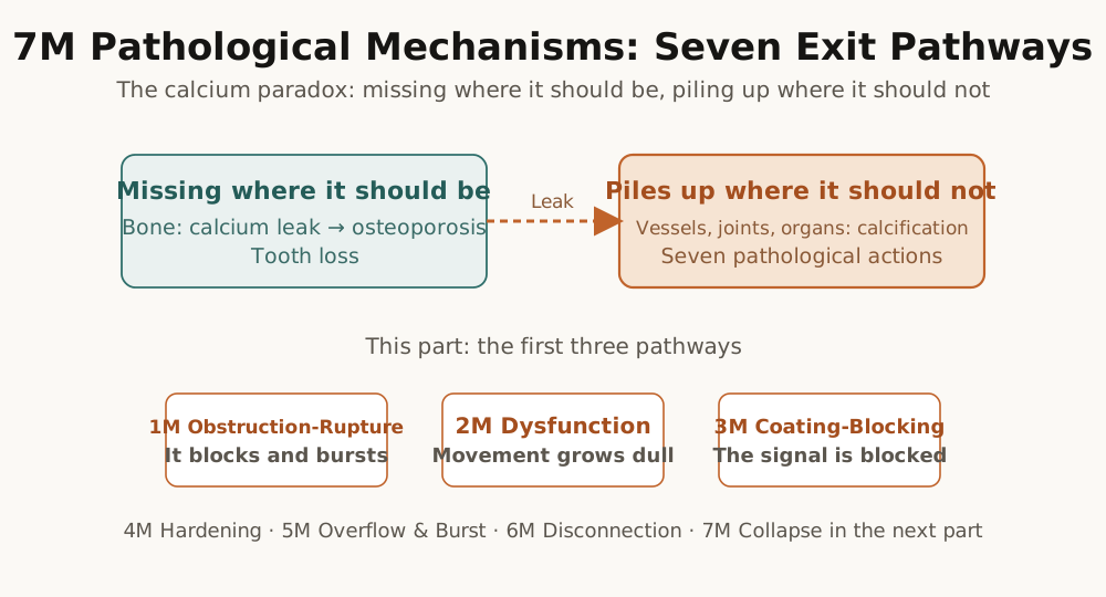
Introduction: Where Does Leaked Calcium Go
We have looked at how the four DIAH triggers (D Deficiency, I Inflammation, A Acidosis, H Hypoxia) cause calcium to leak out of the bones, and how calcium performs the role of an execution code in the process of evolution. We also confirmed that the leaked calcium meets two fates. One is that it activates immune cells and sets off the vicious cycle of inflammation, and the other is that it binds with phosphate and deposits as calcification in the soft tissues.
The answer lies in the seven pathological mechanisms, that is, 7M (Mechanisms). When the calcium that has escaped from the bones reaches the soft tissues, it can cause damage in different ways depending on its location and concentration and the characteristics of the tissue. In some places it blocks a channel, in some places it makes movement dull, and in some places it shuts off secretion.
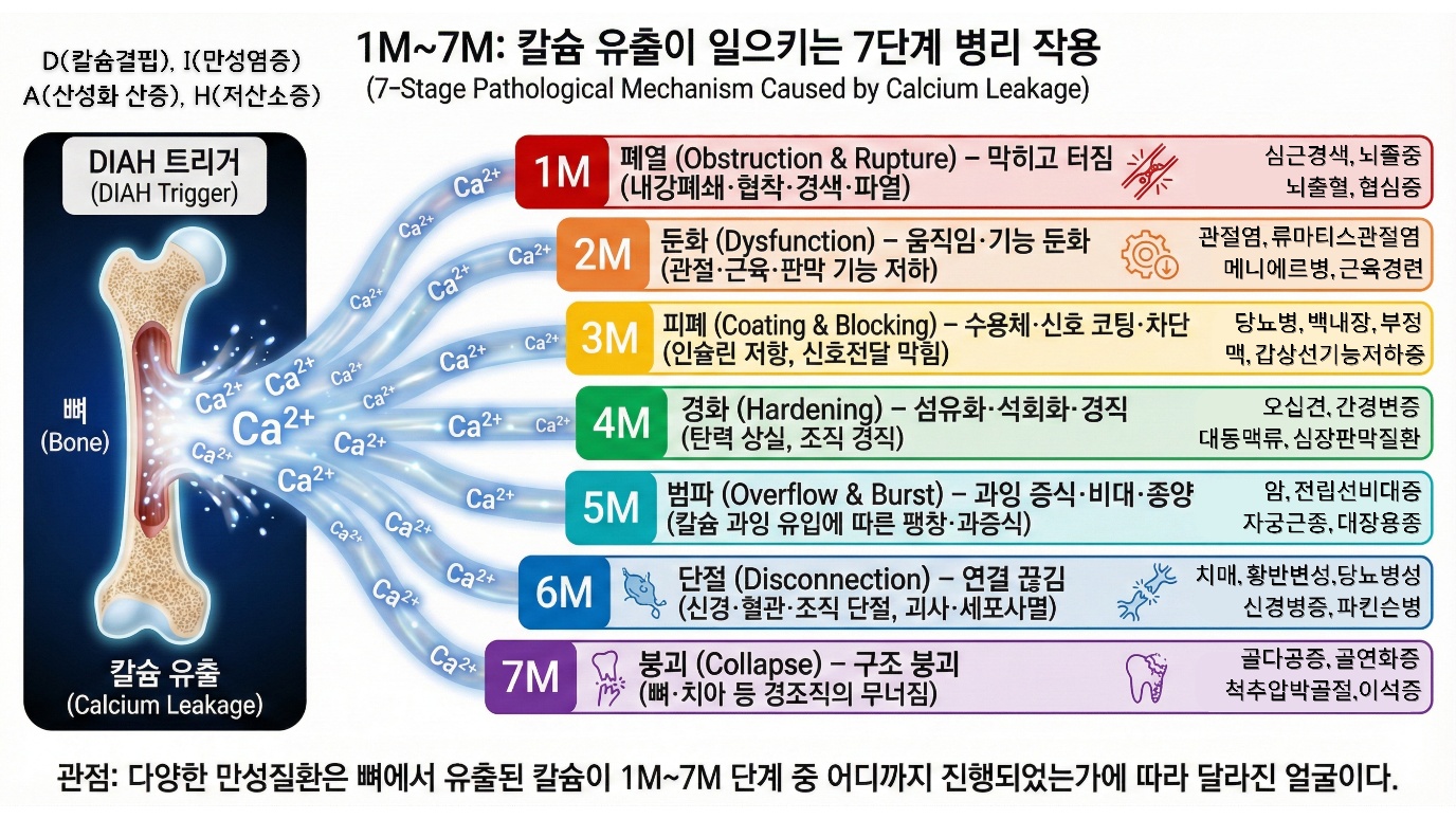
These seven mechanisms are the principal mechanisms that contribute to aging and to the onset of hundreds of chronic diseases.
The Normal Flow of Calcium
In a healthy body, calcium is precisely regulated. When you take in about 1,000 milligrams of calcium a day from food, about 200 to 400 milligrams of this is absorbed in the intestine. The absorption rate is between about 20 and 40 percent, and when vitamin D is sufficient the absorption rate rises, while when it is lacking it falls. The absorbed calcium enters the blood, and the blood calcium concentration is maintained within a very narrow range of about 8.5 to 10.5 mg/dL. If it goes outside this range life is endangered, for if it is too low muscle cramps and cardiac arrhythmia occur, and if it is too high changes in consciousness and cardiac arrest occur. Blood calcium goes to several places. It goes to the bone and is used for bone formation, it goes to the cells and is used for signal transmission, and it goes to the kidneys where some is excreted in the urine, with about 100 to 200 milligrams a day leaving in the urine. Calcium also comes out of the bone, for when osteoclasts resorb bone during the bone remodeling process calcium is released into the blood. In a healthy state, as much is formed as is resorbed and a balance is reached, and overall the amount coming in and the amount going out are balanced so that the calcium reserve of the bone is kept stable. This is calcium homeostasis.
Bone can be likened to a bank. The total assets of the calcium bank are about 1 to 1.2 kilograms. Since 99 percent of the body's calcium is stored in the bone, it is an enormous scale. Deposits and withdrawals occur every day, in which the calcium absorbed from food is deposited and the calcium excreted in urine and feces is withdrawn, while the calcium used in the cells is also a withdrawal. In a healthy state, deposits and withdrawals are balanced and the bank balance is maintained. But in the DIAH state the balance is broken. In the deficiency state, deposits decrease. When calcium intake is insufficient, or absorption is impaired, or vitamin D is lacking, the calcium absorbed in the intestine decreases. But the body must maintain the blood calcium concentration without fail. Even if the blood calcium drops just a little, the parathyroid gland detects it and parathyroid hormone is secreted, and parathyroid hormone is a command to mobilize calcium from the bone. In inflammation, acidosis, and hypoxia as well, calcium efflux from the bone increases. As we examined in detail in Chapter 6, each trigger activates osteoclasts through the RANKL-RANK-OPG system. The result is the same. Deposits decrease and withdrawals increase. The bank balance falls. This is net efflux.
The calcium that has escaped from the bone enters the blood. What happens next?
If things were normal, three pathways would be open. First, return to the bone. This is the pathway in which osteoblasts make new bone and use the calcium again. Second, excretion through the kidneys. When blood calcium rises slightly, excretion in the kidneys increases. Third, use in the cells. This is its use for physiological functions such as signal transmission, muscle contraction, and neurotransmission. But in the DIAH state these pathways do not work properly.
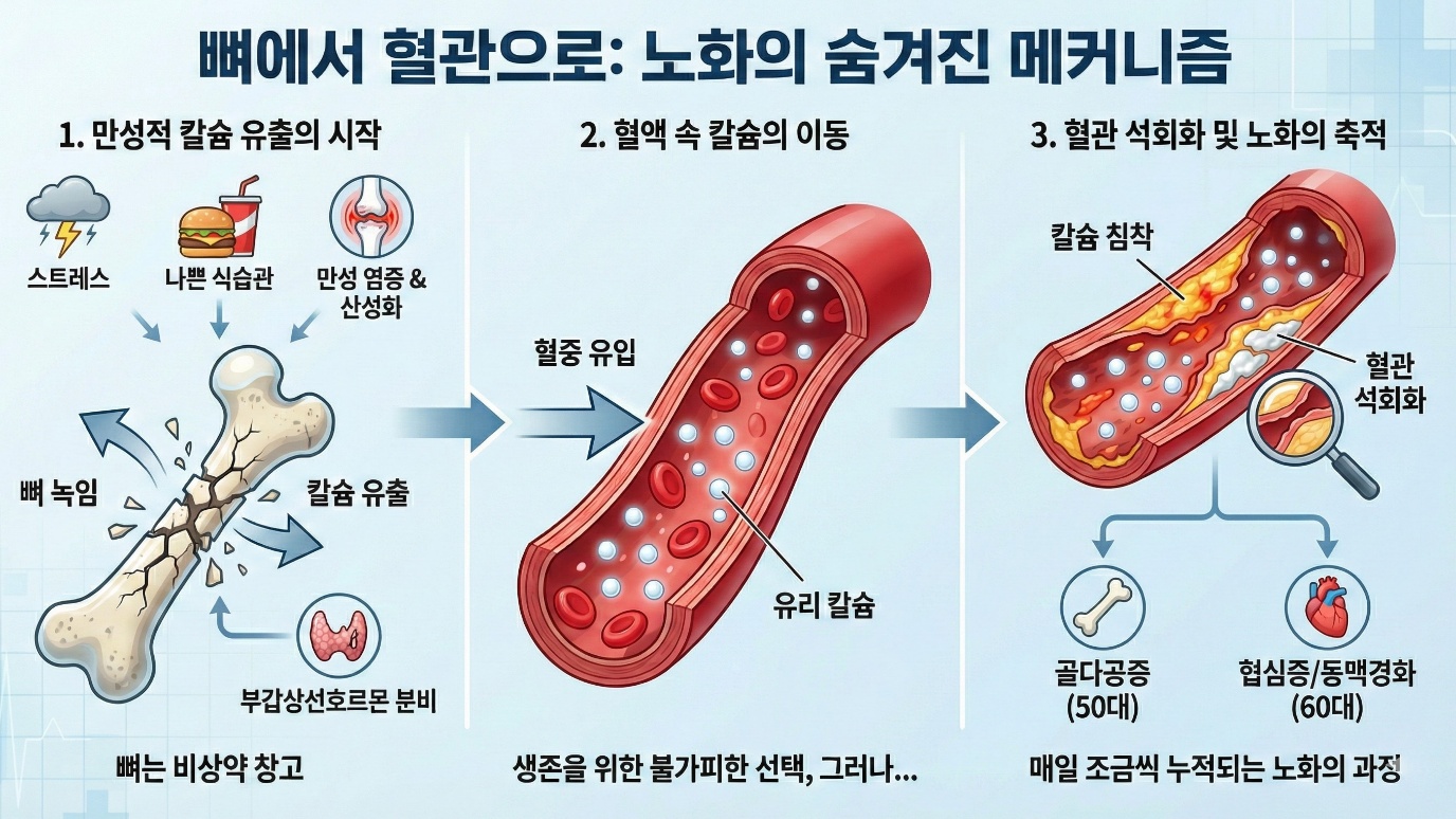
Bone formation is impaired. This is because inflammatory cytokines suppress osteoblast function. The escaped calcium cannot re-enter the bone. Kidney excretion also has its limits. Especially in the case of the elderly, the capacity to excrete calcium is restricted by declining kidney function. As a result the calcium comes to wander through the body. Having come out of the bone but unable to return to the bone, and unable to be fully excreted either. Where does this calcium go?
It piles up in the wrong places. In blood vessels, in joints, in valves, in organs. Calcium deposits in places where it originally should not be. This is called ectopic calcification. "Ectopic" means in a place other than where something should originally be. Ectopic calcification: it is here that 7M begins.
The Calcium Paradox: Absent Where It Should Be Present
In the clinic there is an interesting observation. Vascular calcification is more common in osteoporosis patients. Calcium is lacking in the bone yet calcium has piled up in the blood vessels. According to several epidemiological studies, a tendency is observed in which the lower a person's bone density, the higher the coronary artery calcium score and the greater the risk of cardiovascular events. Aortic calcification accompanies more often in people who have vertebral fractures. Osteoporosis and cardiovascular disease tend to occur at the same time in the same person. This is the calcium paradox. It is the same calcium, yet it is absent from the bone where it should be present, and present in the blood vessels where it should not be. How is such a thing possible?
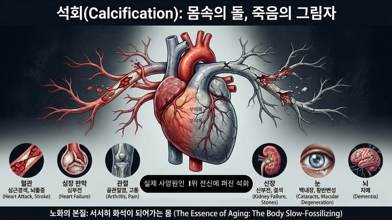
The calcium paradox is in fact not a paradox. It is only two aspects of the same process. Calcium escapes from the bone, and the escaped calcium piles up in the blood vessels: it is a cause-and-effect relationship in which the decrease on one side creates the increase on the other. Looked at more fundamentally, the calcium regulation of bone and of blood vessels is connected to each other. Estrogen is a good example.
Estrogen promotes bone formation and suppresses bone resorption in the bone. At the same time it performs a protective action in the blood vessels as well. Improving endothelial cell function, suppressing inflammation, suppressing calcification. It is, in effect, a hormone that guards both the bone and the blood vessels at once. So what happens when estrogen decreases with menopause?
In the bone, calcium begins to escape. At the same time, vascular protection weakens too. In the end the calcium that has come out of the bone comes to pile up in the blood vessels that have lost their protective shield. Consider vitamin K2. Vitamin K2 activates osteocalcin in the bone and promotes bone formation. At the same time it activates matrix Gla protein in the blood vessels and suppresses calcification. In the healthy years of youth this system works well. Little calcium escapes from the bone, and the vitamin K2-dependent proteins handle it sufficiently. Osteocalcin returns calcium to the bone, and matrix Gla protein blocks vascular calcification.
But when one enters the period of decline the situation changes. As the DIAH triggers are activated, a large amount of calcium leaks from the bone. The problem is that the processing capacity of the vitamin K2 system has its limits. No matter how sufficient the vitamin K2 is, it cannot handle the amount of calcium pouring out. It is like a dam bursting and water pouring out while one bails it out with a bucket. Moreover, with aging the activity of the vitamin K2-related enzymes themselves declines as well. Changes in the gut microbiota reduce vitamin K2 synthesis, and the absorption rate falls too. It is a double problem. The calcium that must be processed increases, while the processing capacity decreases.
The same regulator regulates bone and blood vessels in opposite directions. Putting calcium into the bone and taking calcium out of the blood vessels is the same system. When this system weakens, both are reversed. Calcium leaks from the bone and calcium piles up in the blood vessels. But the more fundamental problem is that, because of DIAH, the very amount of calcium leaking from the bone is excessive. Vitamin K2 supplementation alone cannot stop this flood.
Why is the calcium regulation of bone and blood vessels connected? Because it is evolutionarily efficient. This is explained by the theory of antagonistic pleiotropy that Williams proposed in 1957 and the disposable soma theory that Kirkwood proposed in 1977. It is that a trait advantageous in the reproductive period can become harmful after reproduction. In the reproductive period, the bone must be protected. A sturdy skeleton is advantageous for survival and reproduction.
At the same time, the blood vessels must also be protected. A healthy circulatory system is advantageous for survival and reproduction. It is efficient to protect both with a single regulatory system. There is no need to make a separate system. After reproduction, protection is not needed. The genes have already been passed on. If you weaken a single regulatory system, both are weakened. The bone weakens too and the blood vessels weaken too. It is an efficient exit. The calcium paradox is not a paradox but an evolutionary design.
What does this imply? In the period of decline, calcium supplementation is important above all. One must ease the DIAH triggers and prevent calcium from leaking excessively out of the bone. The heart of that is to supplement sufficiently with calcium whose absorption rate has been verified. If blood calcium is sufficient, parathyroid hormone has no reason to attack the bone. It is blocking deficiency, the first trigger. Vitamin K2 plays a supporting role. It helps place calcium in the right spot at the right time, but it cannot stop the situation in which calcium pours out of the bone in the first place. To respond to the root cause, calcium deficiency must be resolved.
The Seven Pathological Actions That Calcium Leaked from the Bone Causes in the Body
| Mechanism | Korean name | English name | Action | Scope |
|---|---|---|---|---|
| 1M | Pyeyeol | Obstruction & Rupture | It blocks and bursts | calcium deposition -> luminal obstruction, stenosis, infarction, rupture, hemorrhage |
| 2M | Dunhwa | Dysfunction | It grows dull | calcium deposition -> dulling of joint/muscle/valve movement, decline of contraction/relaxation function |
| 3M | Pipye | Coating & Blocking | It is coated and blocked | calcium deposition -> receptor blocking, signal blocking, secretion blocking, insulin resistance |
| 4M | Gyeonghwa | Hardening | It hardens | calcium deposition -> fibrosis, calcification, stiffening, loss of elasticity |
| 5M | Beompa | Overflow & Burst | It overflows and bursts | excessive calcium influx -> cell hyperproliferation, hypertrophy, tumor, expansion, apoptosis |
| 6M | Danjeol | Disconnection | It is cut and severed | calcium deposition -> nerve severance, vascular occlusion, tissue necrosis, apoptosis |
| 7M | Bunggoe | Collapse | It falls apart | calcium deficiency -> structural collapse of hard tissue (bone, teeth) |
1. 1M Obstruction & Rupture: It Blocks and Bursts
Now let us look at each of the 7M. 1M Obstruction & Rupture is when the vascular lumen is blocked, or a blood vessel ruptures, or hemorrhage occurs. Calcification narrows the vascular lumen. Stenosis occurs. If it progresses further it is completely blocked. Infarction occurs. Or the calcified vessel wall weakens and ruptures. Hemorrhage occurs. Cardiovascular disease is the number one cause of death worldwide, accounting for a considerable portion of all deaths. The coronary artery calcium score actually used in cardiology is precisely an index that quantifies the degree of this calcification. According to a review published in the Journal of the American College of Cardiology in 2018, the higher the calcium score, the greatly increased the risk of myocardial infarction and cardiovascular death.
Let us look concretely at the mechanism of vascular calcification. In atherosclerosis, plaque forms on the arterial intima. Plaque is composed of lipids, inflammatory cells, and fibrous tissue. Calcification occurs in the late stage of this process. When macrophages within the plaque die, cell debris remains. This debris becomes the nucleus of calcification. The phospholipids of the dead cell membranes bind with calcium. Small calcium crystals form. This is microcalcification. When microcalcification begins, the vascular smooth muscle cells change. As explained earlier, they convert into an osteoblast-like form. They actively drive calcification forward. The microcalcifications merge and become large calcification. The plaque hardens.
Calcified plaque brings about two results. First, the internal diameter of the blood vessel narrows. Blood flow is restricted. When a coronary artery narrows by 70 percent or more, angina occurs. During exercise or stress, sufficient blood flow is not supplied to the myocardium. Second, the plaque can become unstable. Microcalcification concentrates mechanical stress within the plaque. Force is concentrated between the hard calcium particles and the soft lipid tissue. The plaque becomes prone to rupture. When the plaque ruptures, a thrombus forms. When the lipid core is exposed to the blood, platelets are activated. The coagulation reaction begins. The thrombus blocks the artery. When a coronary artery is blocked it is myocardial infarction. When a cerebral artery is blocked it is cerebral infarction. The tissue dies.
Let us look also at the mechanism of vascular rupture. The arterial wall is composed of three layers. The intima, the media, and the adventitia. In the media there are smooth muscle cells and elastic fibers that give the artery elasticity and strength. When medial calcification occurs, the structure of the arterial wall changes. The elastic fibers are replaced by calcification. The artery hardens. Elasticity disappears. Here there is an important physical principle. Stress concentrates at the interface where soft tissue and a hard mass of calcium meet. When blood pressure rises, the soft tissue stretches but the calcium does not. That boundary is prone to tearing.
An artery that has lost its elasticity is vulnerable to changes in pressure. Every time the heart contracts, a pressure wave is transmitted. A normal artery expands and absorbs the pressure. A calcified artery does not expand. Stress concentrates on the wall. An aneurysm can form. A weak part of the arterial wall bulges out like a balloon. Aortic aneurysm is the most common. When an aneurysm ruptures, massive hemorrhage occurs. When an abdominal aortic aneurysm ruptures, most people die before reaching the hospital. In the brain, rupture of a cerebral aneurysm induces subarachnoid hemorrhage. Rupture of a small artery induces cerebral hemorrhage.
Looked at from an evolutionary perspective, why of all things was calcium mobilized to block and burst blood vessels? First, calcium is already abundant in the blood. It is ready to deposit the moment conditions are right. Second, calcium forms crystals that do not dissolve. Calcium phosphate is almost insoluble in water. Once a crystal forms it does not dissolve easily. It becomes a permanent structure. Sodium salts or potassium salts dissolve well in water, so they cannot play this role. Third, the mechanism for making bone already exists. The molecular machinery by which osteoblasts deposit hydroxyapatite has already evolved. Vascular cells need only borrow this program. There is no need to invent a new mechanism. Fourth, calcification is hard to reverse. Fat deposition can dissolve. Cholesterol can be removed. But calcification can hardly be reversed. Once it progresses it is permanent. As an exit mechanism it is certain.
2. 2M Dysfunction: Movement Grows Dull
2M Dysfunction is when the movement of joints, muscles, and valves grows dull. Contraction and relaxation, opening and closing, do not go smoothly. Let us look at the calcification of joint cartilage. Normal joint cartilage is hyaline cartilage. Chondrocytes are embedded in a matrix made of collagen and proteoglycans. This matrix provides elasticity. It absorbs shock when the joint moves. When calcium deposits in cartilage, chondrocalcinosis occurs. The most common form is calcium pyrophosphate crystal deposition. Chondrocytes produce pyrophosphate. Normally pyrophosphate suppresses calcification. But with aging the metabolism of the chondrocytes changes. The balance of pyrophosphate production and breakdown is broken. Pyrophosphate accumulates in excess. Excess pyrophosphate binds with calcium. Crystals form. Crystals deposit in the cartilage matrix. According to a review published in the New England Journal of Medicine in 2016, from the 60s onward chondrocalcinosis on X-ray increases to roughly 10 percent or more, and after the 80s it is reported to be as high as 20 to 40 percent depending on the study. It is an almost universal phenomenon of aging.
What happens to calcified cartilage? It loses elasticity. Cartilage with crystals embedded in it is hard. It cannot absorb shock. The stress transmitted to the joint increases. The surface grows rough. Crystals protrude on the once-smooth cartilage surface. Friction increases. The cartilage wears down. Inflammation is induced. When crystals fall off into the joint cavity, immune cells respond. Macrophages try to phagocytose the crystals. Inflammatory cytokines are secreted. Acute inflammation occurs. This is pseudogout. Chronically it progresses to osteoarthritis. The cartilage wears away and disappears. Bone strikes against bone. Osteophytes form. Pain occurs. The joint's range of motion decreases. Movement grows dull.
Let us look also at the calcification of the heart valves. The aortic valve is one of the most important valves in the heart. It lies between the left ventricle and the aorta. It is composed of three semilunar cusps. When the heart contracts it opens and blood goes out to the aorta, and when it relaxes it closes and blocks backflow. The aortic valve opens and closes about 3 billion times over a lifetime. It receives enormous mechanical stress. How does valve calcification begin? Mechanical stress concentrates on the aortic-side surface of the valve. The endothelial cells are damaged. Lipids deposit.
Inflammatory cells infiltrate. A process similar to atherosclerosis takes place in the valve. The valvular interstitial cells change. Like vascular smooth muscle cells, they convert into an osteoblast-like form. Calcification progresses. According to a review published in the Journal of the American College of Cardiology in 2012, aortic valve calcification is reported to be about 20 to 30 percent in those aged 65 and over. Of these, in about 3 percent or so it progresses to severe stenosis.
What happens to a calcified valve? It stiffens. The valve that once opened and closed flexibly hardens. It does not open properly. For the left ventricle to push out blood it must create a higher pressure. This is stenosis. A burden falls on the left ventricle. To create a higher pressure the myocardium hypertrophies. At first it is a compensatory response. But if it continues the myocardium grows exhausted. Heart failure occurs.
Once symptoms appear the prognosis is poor. It is known that after symptoms of angina, syncope, and heart failure appear, if left untreated the prognosis worsens sharply. Only valve replacement is a fundamental treatment. It is replacing the dulled valve with a new one. Looked at from an evolutionary perspective, joints and valves have something in common. Both are moving structures. Both must be flexible. Both receive repeated mechanical stress throughout life. Calcium crystals are hard.
When hard crystals become embedded in flexible tissue, flexibility disappears. Movement grows dull. What happens to a dulled individual in the wild? When the joints grow dull it cannot run. It cannot escape predators. It cannot catch prey. When the valves grow dull it cannot withstand intense activity. It grows exhausted in a chase. It fails to flee. Dysfunction is a slower exit pathway than Obstruction & Rupture. One does not die suddenly. One is slowly pushed out of the competition. In the end it arrives at the same result.
3. 3M Coating & Blocking: The Signal Is Blocked
3M Coating & Blocking is when a receptor is blocked and a signal is cut off. When calcium obstructs a receptor pathway, the signal is not transmitted. Insulin resistance is representative. Let us take insulin resistance as an example. Insulin is a hormone secreted from the beta cells of the pancreas. When blood sugar rises after a meal, insulin is secreted. Insulin binds to the insulin receptor of the target cell. The signal transduction pathway is activated. Glucose transporters move to the cell membrane. Glucose enters the cell. Blood sugar falls. Insulin resistance is a state in which this process does not work properly. Even when insulin is secreted, the cell does not respond.
How does calcium take part in insulin resistance? In a normal cell the cytoplasmic calcium concentration is kept very low, at about 100 nanomoles. As we examined in Chapter 1, the cell spends enormous energy to keep calcium low. But in chronic inflammation, obesity, aging, and so on, cytoplasmic calcium rises slightly. From 100 nanomoles to 200 to 300 nanomoles. On the surface it is a small change. But the consequence is large. The raised calcium impairs insulin signal transduction. How? Calcium activates several enzymes. In particular, protein kinases are activated. This enzyme phosphorylates the insulin receptor substrate at a specific site. Normally the insulin receptor phosphorylates the insulin receptor substrate at a different site. Only then is the signal transmitted. But when the wrong site is phosphorylated by calcium, normal phosphorylation is impaired. Signal transduction is blocked. The insulin is there but does not work. The receptor has been blocked. This is Coating & Blocking. A study published in the journal Diabetes in 2006 laid out that this serine phosphorylation mechanism is a core pathway of insulin resistance. In reality, insulin resistance operates through a complex of fat accumulation, inflammation, and genetic factors, and the disturbance of intracellular calcium signaling has also been proposed as one of several contributing factors.
The pancreatic beta cells are affected too. In the pancreas of a type 2 diabetes patient, amylin protein accumulates. Amylin is a hormone secreted together with insulin. Normally it is dissolved. But when it is secreted in excess it aggregates. It forms amyloid. Calcium deposits on this amyloid. Calcified amyloid piles up around the beta cells. The beta cells are surrounded by this deposit and their function is impaired. Insulin secretion decreases. To insulin resistance is added decreased insulin secretion. This is type 2 diabetes. It is not only insulin. Other secretory organs are affected too. Stones form in the salivary glands. Calcium is the main component. The salivary duct is blocked. Saliva secretion is impaired. Dry mouth occurs. Calcification occurs in the pancreas. It is common in chronic pancreatitis. The pancreatic duct is blocked. Digestive enzyme secretion is impaired. Indigestion occurs. Calcification occurs in the thyroid. It is common in thyroid nodules. It can affect thyroid function. When secretion is blocked, the regulation of hormones and enzymes collapses. The coordination of the whole body is broken.
Looked at from an evolutionary perspective, why of all things was calcium mobilized to block signals? Paradoxically, it is because calcium is a signaling molecule. As we examined in Chapter 1, calcium is the core of intracellular signal transduction. When the calcium concentration rises, countless enzymes are activated. This is the signal. But a signal must be temporary. It must switch on and off. Calcium must rise and then fall. What happens when calcium is chronically slightly high? The signal is in a permanently on state. The calcium-dependent enzymes are always activated. The signal system is saturated. It cannot detect a new signal. Because it is already always on. Even when insulin comes it cannot respond. Because the downstream pathway is already blocked. Because calcium is a signaling molecule, a disturbance of calcium homeostasis blocks signal transduction. This is Coating & Blocking.