
This chapter looks at a single phenomenon longer and more deeply than any other chapter in this book. It is the phenomenon of cancer. A reader who has read this book this far will not expect to learn the pathology of cancer anew in this chapter. The purpose of this chapter lies elsewhere. It is the work of confirming how the single grammar of gradient is revealed even in cancer, the most extreme case, and, through that confirmation, of the reader gaining one line of sight from which to view this vast phenomenon placed before them a little differently.
The single sentence that runs through this entire chapter is this book's second single-sentence declaration. "Cancer is not the rebellion of cells but the result of an environment." This single sentence is the spine of all the discussion that follows. But this declaration does not mean that it explains every kind of cancer. Before entering the main discussion, we must first clearly establish the boundaries of this chapter.
The Boundary of This Chapter: Which Cancers Does It Address
This chapter addresses adult solid cancers that arrive with aging. Colorectal cancer, breast cancer, prostate cancer, stomach cancer, hepatocellular carcinoma, pancreatic cancer, lung cancer, kidney cancer, endometrial cancer, bladder cancer, and esophageal cancer belong here. These cancers commonly rise steeply in incidence after the forties and grow on top of the long-term accumulation of environmental conditions, namely hypoxia, acidosis, inflammation, and microvascular abnormality. It is precisely these adult solid cancers to which the grammar of gradient that this book has built up earlier applies most vividly.
There are cancers the language of this chapter does not reach. Childhood cancers begin from abnormalities in the embryonic developmental process or from early-life mutations, and they do not pass through the long-term environmental accumulation pathway this chapter addresses. The same is true of hereditary cancer syndromes in which a germline mutation is the primary cause, such as BRCA1-related breast and ovarian cancer, Lynch syndrome-related colorectal cancer, and Li-Fraumeni syndrome. Cancers in which infection is the primary cause, such as HPV-related cervical cancer, the virus-driven portion of hepatitis B and C virus-related liver cancer, and HTLV-1-related adult T-cell leukemia, are also outside the domain of this chapter. Most blood cancers, including acute leukemia, are primarily driven by somatic mutations of hematopoietic cells, and occupational cancers in which a single high-intensity exposure is the primary cause, such as asbestos mesothelioma, likewise have a separate pathway.
The reason for noting this boundary is simple. The language of gradient has its strongest explanatory power in cancers where the environment is the primary stage. If this language is forcibly extended to cancers outside that, its explanatory power decreases and misunderstanding increases. All discussion in this chapter presupposes the adult solid cancers enumerated in the first paragraph. The goal is to show what this chapter can say within this boundary, and how consistently that saying connects with the previous chapters.
But let me add one thing. Cancers outside the domain of this chapter also pass through the same bottleneck as aging-associated cancers in their progression pathway after onset. Whether a hereditary cancer, a virus-induced cancer, or a cancer that began from occupational exposure, from the moment the tumor grows beyond a certain size it enters the common microenvironment of hypoxia, acidosis, inflammation, and microvascular abnormality, and that environment determines a considerable part of the speed of progression. This chapter does not integrate the causes of onset, but it shows where the pathways of progression meet as one. The beginnings differ, but the door they pass through is the same. This is the second axis of this chapter. This observation of convergence is noted once more at the appropriate place in the main text.
The Places Conquered and the Places Not Yet
Half a century has passed since U.S. President Nixon declared the "War on Cancer" in 1971. In this half-century, humanity has invested enormous resources in cancer research. The annual budget of the U.S. National Cancer Institute (NCI) exceeds 7 billion dollars as of the 2020s, and the number of clinical trials worldwide expanded to the point that at one time cancer alone accounted for half of all pharmaceutical clinical trials. This investment has borne clear fruit. Science and technology descended to the molecular unit of the phenomenon of cancer, numerous targeted therapies and immunotherapies were introduced into the clinic, and the accuracy of imaging diagnosis and early detection became more refined than a generation ago beyond comparison.
Seen concretely, the fruits are as follows. According to the annual report of the American Cancer Society (ACS), between 1991 and 2021 the age-adjusted cancer mortality rate decreased by about 33%, and thanks to that, it is estimated that about 4.1 million lives were additionally saved during that period. The five-year survival rate of certain cancer types has improved dramatically. Childhood acute lymphoblastic leukemia was virtually incurable in the 1960s but now shows a cure rate close to 90%, and chronic myeloid leukemia has become a virtually long-term manageable disease since imatinib (Gleevec). The five-year relative survival rate of breast cancer has entered the 90s thanks to early-detection screening systems and hormone therapy, and colorectal cancer and prostate cancer show the same trend.
But the other half of the landscape is still dark. There are domains where a half-century of investment still meets a wall. The five-year survival rate of pancreatic cancer has barely moved from around 10% for decades, and the survival rates of the advanced stages of hepatocellular carcinoma and biliary tract cancer are in a similar place. Lung cancer's overall survival rate is improving thanks to targeted therapy and immunotherapy, but it is still the number-one cause of cancer death, and long-term survival at the advanced stage is limited. Ovarian cancer, esophageal cancer, and glioblastoma (malignant brain tumor) are in a similar place. And most decisively, for any cancer type, it is difficult to say that the long-term survival rate after distant metastasis has occurred has changed dramatically compared with 30 years ago. Excluding cases where long-term survival of metastatic cancer in specific molecular subtypes has noticeably improved (HER2-positive breast cancer, BRAF-mutant melanoma, etc.), cure after metastasis is still the exception.
In summary, humanity has walked a half-century toward the vast phenomenon of cancer, and the places its steps have reached and the places they have not are divided. In cancers that can be caught by early detection, cancers in which a single molecular target plays a decisive role, cancers with strong hormone dependence, and subtypes the immune system already recognizes, there has been dazzling progress. Conversely, in cancers with a complex microenvironment, high molecular heterogeneity, an inevitably late point of detection, and metastasis already progressed, progress has been limited. This asymmetry is the starting point of this entire chapter. Why is progress easy in some cancers and so difficult in others? If this question can be viewed in the language of gradient, we can gain at least one layer of interpretation.
The Reason It Is Reborn Even After You Defeat the Cell
The dominant strategy of modern oncology is to target the tumor cell itself. Cytotoxic anticancer drugs interfere with the DNA replication of rapidly dividing cells, targeted therapies inhibit the activity of a specific mutant protein, and immunotherapies help the immune system recognize a specific protein on the surface of the tumor cell. These strategies work. Many of the tumor cells respond to treatment, the lesion shrinks on imaging, and tumor markers go down. But in many cancer types, especially at the advanced stage, after the first few months or few years of response, the cancer comes back. A differently shaped clone grows, lesions appear in other organs, and the same drug no longer works. This is the greatest clinical frustration of modern oncology.
The existing grammar explains the reason for this frustration as resistance and heterogeneity. Within a tumor, cells with various genetic mutations are mixed from the start, and treatment kills the susceptible cells among them, but the cells that are not susceptible or that adapt survive and grow again. This explanation is accurate at the molecular level. But it naturally summons one question. Why are such resistant clones born so well, so repeatedly, and so identically in so many patients?
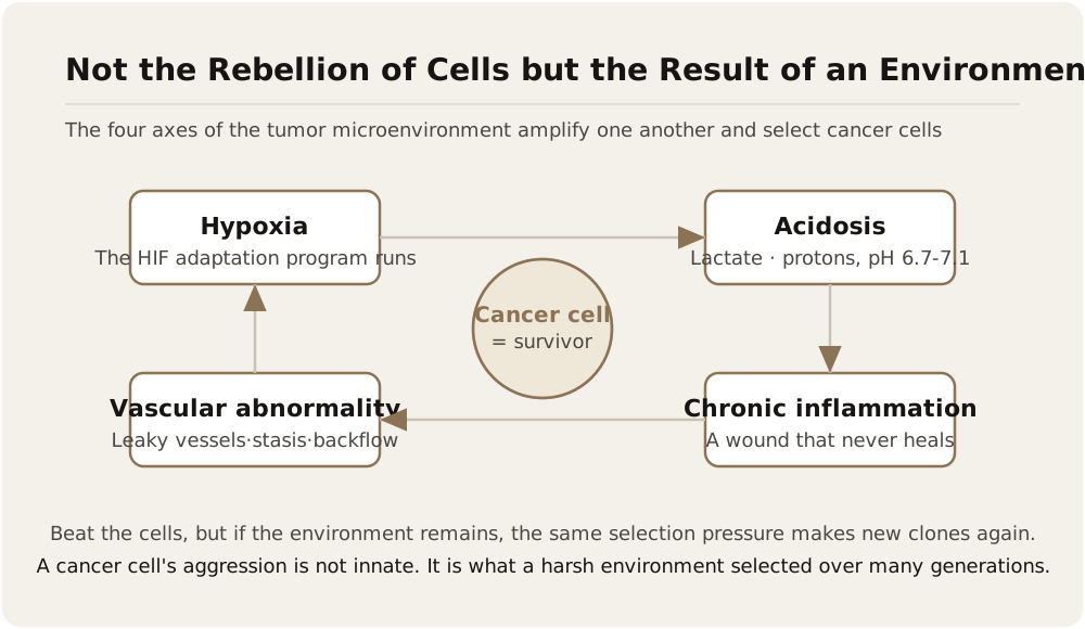
From the perspective of this book, the answer is the environment. The microenvironment of the tissue in which the tumor takes root is a harsh ecosystem in which hypoxia, acidosis, chronic inflammation, and abnormal vascular structure are tangled together. For a cell to survive in such an environment, it must turn on an adaptation program that ordinary cells do not use. It must change glucose metabolism, summon new vessels, evade immune surveillance, push out neighboring cells, and sometimes even acquire the ability to move across tissue boundaries. The cell that survives at each step of this adaptation is precisely what we call a "cancer cell." In other words, the aggressiveness of a cancer cell is not a property inherent in the original normal cell, but the result that a harsh environment has selected out through many generations of adaptation.
If one accepts this perspective, the riddle of resistance and heterogeneity looks different. So long as the environment remains as it is, even if we kill a specific clone, the environment produces a new clone under the same selection pressure. Even if we defeat the cell, the environment makes new cells grow. The tumor does not "possess" resistance; the environment "produces" resistance. This shift of view is the core of this chapter. To fight cancer, one must see not only the cell but the entire ground on which the cell is placed.
In the earlier chapters of this book, we viewed the cell not as cause but as response. We said that the cellular abnormality of chronic disease is not a breakdown of the cell itself but a rational adaptation to the long-term decline of the microenvironment. Cancer is the case in which this same principle is revealed most extremely. A state in which a normal cell, after repeated adaptation under decades of pressure from hypoxia, acidosis, inflammation, and gradient collapse, has lost control of proliferation and movement: this is cancer as this book understands it. The cancer cell is not a rebel army but a survivor who lasted to the end in an environmental state of emergency that continued far too long.
The Microenvironment: Hypoxia, Acidosis, Inflammation, Gradient Collapse
The place where a tumor grows is a distinctive ecosystem. Since Judah Folkman proposed the angiogenesis-dependence of tumors in the 1970s, the research of the past half-century has revealed this ecosystem in considerable detail (Folkman, 1971, N Engl J Med). The constituent elements of the tumor microenvironment are broadly divided into four. First, a new vascular network created by abnormally proliferated endothelial cells. Second, the stroma formed by activated fibroblasts and immune cells. Third, the metabolic products and signaling molecules secreted by the tumor cells themselves. Fourth, the physicochemical conditions of hypoxia, acidosis, inflammation, and gradient collapse that all these elements create as they tangle together.
Of these, the most central in the grammar of gradient is the fourth. In healthy tissue, a cell always has a microvessel within 20 micrometers. This distance is the limit at which oxygen and glucose can reach the cell by diffusion alone, and within this limit the cell maintains the balance of supply and discharge. As a tumor grows, angiogenesis occurs, but the newly formed vessels differ in structure from normal vessels. The tube walls are incomplete, the distribution of branches is irregular, and blood flow stagnates and refluxes. As a result, hypoxic regions 50 to 150 micrometers away arise within the tumor, and these regions are placed in a state of chronic hypoxia, acidosis, and waste stagnation.
Hypoxia is the central trigger of this ecosystem. To respond to hypoxia, the cell stabilizes a transcription factor called hypoxia-inducible factor (HIF-1α), and this factor turns on hundreds of genes simultaneously. The list of genes HIF-1α turns on almost coincides with the features possessed by tumor cells. VEGF, which induces angiogenesis; GLUT1, which increases glucose uptake; the mass production of glycolytic enzymes; pH-regulating proteins; and the protein-degrading enzymes that aid cell movement. In other words, many of the characteristics of the tumor cell are the result of the HIF-mediated adaptation program to the environmental condition of hypoxia (Semenza, 2012, Cell).
Acidosis is layered on top of this. When, in a hypoxic state, the cell depends on glycolysis, breaking glucose down only to lactate, lactate and hydrogen ions pour out of the cell. It is not uncommon for the interstitial pH of tumor tissue to fall from the 7.4 of normal tissue to 6.7 to 7.1. This acidic environment is unfavorable to normal cells but provides a relative advantage to tumor cells that have completed various adaptations. The acidic environment lowers the function of adjacent normal cells, suppresses the activity of immune cells, and increases the activity of matrix-degrading enzymes, aiding invasion and metastasis (Corbet & Feron, 2017, Nat Rev Cancer).
Chronic inflammation is the fourth axis of the tumor environment. The macrophages, neutrophils, regulatory T cells, and activated fibroblasts that gather at the tumor continuously secrete inflammatory cytokines, and this environment operates like an infinitely prolonged healing process that has not received the termination signal of normal wound healing. The intuition of the 19th-century pathologist Virchow, who said "a tumor is a wound that does not heal," has, in effect, been confirmed at the molecular level by the research of recent decades. Chronic inflammation causes DNA damage, promotes angiogenesis, and supports the survival and proliferation of tumor cells (Coussens & Werb, 2002, Nature).
These four axes do not operate separately. Hypoxia summons inflammation, inflammation worsens acidosis, acidosis fixes vascular abnormality in place, and vascular abnormality in turn deepens hypoxia. The DIAH square that this book earlier pointed to as the upstream cause of chronic disease is circulating as it is within the tumor microenvironment as well. In fact, the microvascular calcification, basement membrane thickening, interstitial fibrosis, and chronic low-grade inflammation observed in the tissue of a chronic-disease patient are physicochemically continuous with the background environment of the cancer that arises in the same patient. That the incidence of certain cancers is high in people with chronic disease is not a coincidence but closer to the sharing of the same soil.
In summary, the tumor microenvironment is an extreme form of tissue whose gradient has severely collapsed. Both supply and discharge break down, the cell switches to survival mode, and as that survival mode becomes prolonged, control of proliferation and movement comes undone. From this view, cancer is not the betrayal of normal cells but the landscape reached at the end of a gradient collapse that lasted far too long.
The Biology of Overgrowth: The Calcium Signal and Overproliferation
In Chapter 9, we saw the seven damage patterns, the 7M, in which dual blockade surfaces organ by organ. The fifth of them is Overgrowth (氾破). It is the phenomenon in which cells overproliferate or become enlarged. Cancer is the most extreme expression of this Overgrowth. And the molecular central axis of Overgrowth is precisely calcium, which this book has pointed to as the medium from the very beginning.
In a normal cell, the calcium concentration across the inside and outside of the cell maintains a gradient of about ten-thousand-fold. This gradient is a state kept by the calcium pumps and ion exchangers of the cell membrane spending energy, and when the cell receives an external signal, this gradient opens for a very brief instant, so that the calcium concentration inside the cell rises momentarily and then quickly returns to its place. This "momentarily rising and falling" calcium wave serves as the switch for cell division, movement, differentiation, and death. A healthy cell strictly controls the amplitude and duration of this wave.
In a cancer cell, this control comes undone. When the calcium pump function of the cell membrane declines in a state of hypoxia and acidosis, the calcium inside the cell stays at a chronically high level, and the expression of the calcium channels themselves is also reconfigured. In particular, reports have accumulated that in various cancer types the expression of calcium channels such as TRPV6, TRPC6, and ORAI1 increases, and the expression of endoplasmic reticulum calcium pumps such as SERCA decreases (Monteith et al., 2017, Nat Rev Cancer). As a result, the activity of calcium-dependent transcription factors (NFAT, CREB, NF-κB) is placed at a chronically high state over the long term, and these factors chronically turn on genes related to cell proliferation and survival.
Hepatocellular carcinoma research is one of the cases that shows this structure most vividly. A 2020 review by the Cui team published in the journal Cancers (Cui et al., 2020, Cancers) established that the calcium signal is a central hub that simultaneously regulates the proliferation, movement, angiogenesis, and apoptosis resistance of liver cancer cells. The liver is an organ in which the pathway leading through chronic hepatitis, fatty liver, and cirrhosis to hepatocellular carcinoma is very well defined, and at every step of this pathway chronic hypoxia, inflammation, and acidosis accumulate. And the place that accumulation finally reaches is the collapse of calcium homeostasis and the activation of the Overgrowth program. This pathway overlaps almost one-to-one with the five stages of accumulation of determinants → trigger → dual blockade → manifestation → collapse that this book has drawn earlier.
The same molecule is at the center of myocardial hypertrophy as well. According to what the Molkentin team reported in Cell in 1998, calcineurin, a calcium-dependent phosphatase, directly turns on the genetic program that induces the hypertrophy of cardiomyocytes (Molkentin et al., 1998, Cell). Myocardial hypertrophy is not cancer, but it is a typical case of Overgrowth in which the cell abnormally enlarges, and the fact that its key switch is calcium suggests that the pattern of Overgrowth is a universal mechanism that does not discriminate by organ. Studies have accumulated that reconfiguration of calcium channel expression correlates with prognosis in breast cancer, colorectal cancer, prostate cancer, and lung cancer as well.
What this observation means is simple. Cancer is not a state in which the molecular machinery of a normal cell has broken down, but a state in which that machinery is "turned on far too long" in the wrong way in the wrong environment. The calcium wave rings endlessly, the proliferation switch does not turn off, and the survival signal is prolonged. Because the root of this prolongation lies in the environment outside the cell (hypoxia, acidosis, inflammation, gradient collapse), turning off a single molecule inside the cell does not stop this prolongation. It is because, if the environment remains as it is, another molecule takes over the same role. The theoretical basis of the environment-centered intervention this book speaks of lies here.
The Warburg Effect: The Metabolic Shift Is Not the Cause but an Adaptation
In the 1920s, the German biochemist Otto Warburg discovered the fact that tumor cells metabolize glucose by glycolysis alone and put out large amounts of lactate even when oxygen is sufficiently present. He pointed to this phenomenon as the primary cause of the tumor and argued that a primary abnormality of mitochondrial function creates cancer. This hypothesis was highly influential in its day, but as the era of molecular biology opened, the primary cause of cancer was replaced by the view that it was not the mitochondria but the genes. The Warburg effect remained as a phenomenon but retreated to the back seat as a cause.
The research of recent decades is overturning this structure once more. The interpretation has become dominant that the Warburg effect arises not because of a primary breakdown of the mitochondria, but as a rational adaptation to the environmental conditions in which the tumor cell is placed. In a hypoxic environment, the cell cannot fully rely on the oxygen-dependent electron transport chain and must raise the speed of glycolysis to process sugar quickly. At the same time, a rapidly dividing cell needs, in addition to energy, large amounts of the carbon skeletons and reducing power to make new cells, and the intermediate products of glycolysis supply both of these (Vander Heiden et al., 2009, Science).
This reinterpretation naturally meshes with the framework of this book. The metabolic shift is not a breakdown inside the cell but a response to the environment. When the environment is placed for a long time in a state of hypoxia, acidosis, and nutritional imbalance, the cell readjusts its metabolism to survive in this state. That readjustment is precisely the Warburg effect, and the Warburg effect in turn strengthens the acidosis of the surrounding tissue and makes the environment harsher. Acidosis lowers the function of adjacent normal cells and suppresses immunity, consequently raising the relative competitiveness of the tumor cells. The bidirectional feedback in which the environment changes the cell and the changed cell changes the environment again operates here as well.
The clinical meaning of the Warburg effect is two-sided. On one hand it becomes a diagnostic tool. The fluorodeoxyglucose (FDG) used in positron emission tomography (PET) uses the property of tumor cells absorbing glucose in large amounts to image the location and metabolic activity of the tumor. On the other hand it becomes a target of treatment. Cancer-metabolism target drugs such as glycolytic enzymes, lactate transporters, and fatty acid synthesis pathways are being developed, and some are at the clinical trial stage. But here the problem of the environment returns again. Even if a specific metabolic enzyme is inhibited, so long as the environment maintains a state of hypoxia and acidosis, the cell finds another metabolic detour. If the environment does not change, the cell opens a new path and continues its survival.
This far is the answer to the first half of this chapter, that is, "why is cancer the result of an environment." The four axes of the microenvironment select the cell, the calcium signal of Overgrowth turns on proliferation, and even the Warburg metabolism was an adaptation to the environment. In the following part 2, we look at the place where this perspective leads to practice, that is, how metastasis spreads along the gradient, and by what clinical evidence interventions that change the environment are supported.
This article is part (1/2) of the two-part series covering Chapter 10 of The Universal Law: Gradient. The references are consolidated in part (2/2). This article is an educational explanation of theory, not medical advice for the diagnosis, treatment, or prescription of any specific condition. Please consult a medical professional regarding any health-related decisions.