
This piece is the front part of Chapter 7 of Stones in the Body: More Frightening Than Cancer (Yoon Jong-won). It is a narrative containing the author's academic hypothesis, and the body, figures, and citations follow the manuscript as written.
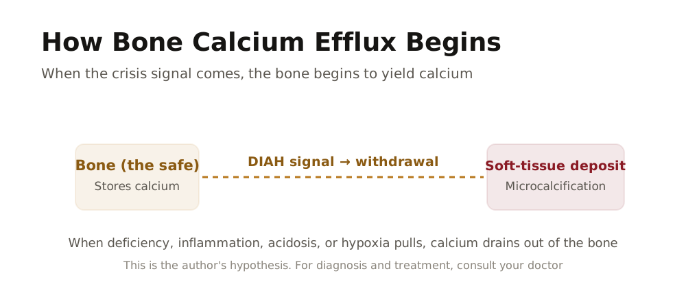
In the previous chapter, through the DIAH Four-Gate Funnel Theory, we examined in detail "what" the factors are that open the bone calcium efflux gate.
We also confirmed that countless factors such as stress, lack of exercise, overeating, drinking, falls, and infection converge into the four gates of D (Deficiency), I (Inflammation), A (Acidosis), and H (Hypoxia), and that this flow is systematized into first order, second order, and third order.
Now the question deepens one step further, from the moment the bone calcium efflux gate opens, in what order and with which triggers turning on does calcium actually begin to leak out inside the body? When D (Deficiency) arises, what alarm does the body sound; when I (Inflammation) lasts long, in which direction does the bone tilt; the more A (Acidosis) accumulates, why does the bone give out calcium like a buffer; and when H (Hypoxia) strikes, in what way does the cell respond and shake the flow of calcium?
This is the second stage of the DIAH-7M Aging and Chronic Disease Pathway System, the domain of ② Start. And because this stage is the most important branch point at which aging and chronic disease "begin in earnest," even if you are not sick now, knowing in advance where things begin to shake becomes the most realistic starting line from which you can design a healthy old age.
Until now, modern medicine has carried a structural limitation: it explained only the factors, symptoms, and final results of disease in isolation, while leaving as a black box, with the process omitted, the question of what choices are actually repeated inside the body in between and through what pathway disease is made.
In this chapter, we will open exactly that black box and look at the process of how aging and chronic disease, as various factors pile up, set off through a certain "start trigger."
[Table] The Five Stages of the DIAH-7M Aging and Chronic Disease Pathway System
| Stage | English | Theory Name | Abbreviation |
|---|---|---|---|
| ① Factor | Factor | DIAH Four-Gate Funnel Theory | DIAH-FG |
| ② Start | Start | DIAH Trigger Bone Calcium Efflux Theory | DIAH-BCO |
| ③ Lockdown | Lockdown | Calcium Absorption and Microcalcification Double Lockdown Theory | CAM-DLT |
| ④ Manifestation | Manifestation | Calcification 7M Pathological Manifestation Theory | Ca-7M |
| ⑤ Result | Result | 7M Life System Collapse | 7M-A |

This chapter deals with that second one, ② Start.
We look at the biochemical mechanism that forcibly draws calcium out of the bone when the four trigger gates open. In particular, you will come to understand how the master key called PTH (parathyroid hormone) opens the safe of the bone, and how the four DIAH triggers connect to one another to form a vicious cycle.
Let us examine this in detail.
Our body has an emergency system to protect life. When the calcium concentration in the blood drops, when inflammation occurs, when the body becomes acidic, or when oxygen runs short, our body immediately opens the emergency safe called bone. This is a survival instinct that has evolved over hundreds of millions of years, and in the short term it is a perfect system that saves life. The reason is that it is the four gates that lead to death.
The problem is that this emergency system has become a daily routine for modern people. As chronic insufficiency of calcium absorption, ceaseless inflammation, acidified eating habits, and the hypoxic state caused by lack of exercise repeat day after day, calcium is pouring out of the bone. When emergency dispatch becomes an everyday event, the emergency safe runs dry, and the calcium that has poured out, unable to find its place, begins to destroy our body.
In this chapter we will look one by one at the four core triggers that forcibly take calcium away from the bone, namely DIAH.
The first is D (Deficiency), where the deficiency of calcium, minerals, vitamin D, and so on is the most direct and powerful trigger.
The second is I (Inflammation), where the immune response consumes the bone.
The third is A (Acidosis), where the bone is sacrificed to neutralize acid.
The fourth is H (Hypoxia), where calcium is excessively consumed for cell survival.
■ D (Deficiency) Subclassification
| Abbreviation | English | Korean | Action | Description |
|---|---|---|---|---|
| D | Deficiency | Deficiency | Bone calcium fills the shortfall | Deficiency of calcium, minerals, vitamin D, and so on → replenished by breaking down calcium from the bone → blood → supplied to various cells ※ Common note: D (Deficiency) is the representative pathway that can directly create a "drop" in blood calcium, while I (Inflammation), A (Acidosis), and H (Hypoxia), depending on the situation, shake the "consumption, distribution, and ionization (availability)" of calcium and, as a result, trigger or accelerate the D pathway (PTH bone mobilization) so that it operates more frequently. |
| I | Inflammation | Inflammation | Calcium is mobilized to quell inflammation | Large amounts of calcium consumed in the inflammatory response → replenished by breaking down calcium from the bone → blood → concentrated at the inflammation site |
| A | Acidosis | Acidosis | Bone is sacrificed to neutralize acid | Alkaline calcium used to neutralize acid → replenished by breaking down calcium from the bone → blood → whole-body pH balance |
| H | Hypoxia | Hypoxia | With oxygen shortage, calcium flows excessively into the cell | Cellular oxygen shortage → ATP decrease lowers Ca²⁺ pump/regulatory function → intracellular calcium overload (damage and death signal) → calcium demand and distribution are shaken during tissue damage and recovery → as a result, when blood calcium "availability" becomes unstable, the D pathway (PTH bone mobilization) is easily called upon → when repeated, a vicious cycle |
D Trigger: Deficiency - Bone Calcium Fills the Shortfall

Blood Calcium: The Absolute Power of 0.1%
The distribution of calcium in our body is extremely unbalanced. 99% of all calcium is stored in the bone, and only the remaining 1% exists in the blood and cells. And even within this 1%, the one that actually performs a physiological function is ionized calcium (Ca²⁺). Only about half of the calcium in the blood exists in the ionized state, and this ionized calcium directly governs our life.

In an adult's blood, only about 0.5g of calcium is circulating. This is a weight slightly lighter than a single one-won coin.
But what this small amount of calcium does is enormous. It makes the heart beat, makes the nerves transmit signals, makes the muscles contract, and makes the blood clot. Calcium is involved in every process by which hormones are secreted and enzymes operate.
According to calcium metabolism research by the U.S. National Institutes of Health, the blood calcium concentration must be precisely maintained within the very narrow range of 8.5 to 10.5 mg/dL.
If it goes outside this range, life is immediately threatened. When blood calcium drops below 7 mg/dL, muscle cramps and numbness in the hands and feet begin; when it falls below 6 mg/dL, cardiac arrhythmia can occur; and below 5 mg/dL, death from cardiac arrest can occur.
Conversely, when it rises above 12 mg/dL, nerve function declines, and when it exceeds 15 mg/dL, clouding of consciousness and kidney damage occur.
Parathyroid Hormone: The Absolute Authority of the Bone Bank

To maintain this narrow range, an elaborate surveillance system operates in our body. At its center is parathyroid hormone (PTH, Parathyroid Hormone).
PTH is a hormone secreted from the parathyroid glands, which are the size of a grain of rice, in the neck; it monitors the blood calcium concentration 24 hours a day, and the moment it drops even slightly, it immediately issues a withdrawal order to the bone.

|According to research published by a U.S. Harvard Medical School research team, the calcium-sensing receptor (CaSR) on the surface of parathyroid cells detects the blood calcium concentration in real time and captures even very subtle changes. This receptor operates like an ultra-precise sensor, and the moment blood ionized calcium approaches the lower limit of the normal range, it immediately increases PTH secretion.
When PTH is secreted, three actions occur simultaneously.
First, it activates the osteoclasts of the bone to break down the bone and release calcium into the blood. Second, it increases calcium reabsorption in the kidney to prevent calcium from leaving through the urine. Third, it activates vitamin D in the kidney to increase calcium absorption in the intestine.
For reference, an intermittent rise in PTH can actually promote bone formation, but the problem is when a chronically high PTH state persists. In this case, osteoclast activity overwhelms osteoblast activity, and bone loss proceeds.

The problem is that among these three actions, the fastest and most effective is the first, namely drawing calcium out of the bone.
Absorbing calcium in the intestine takes several hours, and increasing reabsorption in the kidney is not immediate either. But releasing calcium from the bone happens within a few minutes. In an urgent situation where survival is at stake, our body chooses the fastest method, and that is precisely opening the bone.
The Pathway by Which Calcium Dissolves Out of the Bone: The Double Attack of Parathyroid Hormone
The way PTH draws calcium out of the bone is divided into two stages. One is a fast response, and the other is a sustained response.
1. First stage: rapid calcium release (minutes to several hours)
When PTH is secreted, it first acts on the osteoblasts and osteocytes on the surface of the bone. These cells serve as pumps that quickly release the calcium on the bone surface into the blood. Because the surface area of the bone is very large, through this process alone a considerable amount of calcium can move into the blood within a few minutes.
2. Second stage: osteoclast activation (several hours to several days)
PTH stimulates osteoblasts to produce a protein called RANKL. RANKL binds to the RANK receptor on the surface of osteoclasts and activates the osteoclasts. Activated osteoclasts cling to the bone and secrete acid to dissolve the mineral component of the bone, and secrete protein-degrading enzymes to break down the structure of the bone.

This process is slower than the first stage but far more powerful. A single osteoclast, active over 2 to 3 weeks, can dissolve bone tissue hundreds of times larger than its own size.
In a normal state, osteoblasts make new bone at the spot where osteoclasts dissolved the bone, maintaining balance, but when PTH is secreted excessively, this balance tilts toward bone loss.
Situations in Which the D Trigger Operates
The reasons blood calcium drops are more varied and everyday than one might think. The most common causes are insufficient calcium intake and absorption disorders. According to a survey by the Korean Nutrition Society, the average calcium intake of many Koreans is at a level that falls short of the recommended amount.
In particular, because of eating habits with little intake of high-calcium foods, calcium intake tends to be chronically insufficient. This is the D-NT (nutritional deficiency) trigger.

To make matters worse, there are many cases in which the calcium that is consumed is not properly absorbed. When vitamin D is deficient, the calcium absorption rate in the intestine drops sharply. According to domestic and international research, a considerable number of Koreans are in a state of vitamin D insufficiency or deficiency, suffering the double hardship in which even the calcium they consume is not properly absorbed.
This is the D-VD (vitamin D deficiency) trigger. Also, as one ages, the intestine's ability to absorb calcium decreases, and in menopausal women, calcium absorption becomes even more difficult due to the decrease in estrogen.
To this are added factors that excessively consume or excrete calcium.
Lack of exercise also activates the D trigger. Bone is maintained only when it is stimulated. When you walk, run, or lift heavy things, pressure is applied to the bone, and this pressure sends the bone cells the signal "there is stimulus, so maintain the bone." But when you do not move, this signal is cut off.
When the signal is cut off, there is no reason to maintain the bone, osteoclasts are activated, and the bone begins to dissolve. This is the D-PH (physical stimulus deficiency) trigger. According to NASA research, when an astronaut spends 6 months in a weightless environment, bone density decreases considerably (on the order of 10 to 20% depending on study conditions), and there are reports that even on Earth, if one merely lies in bed, bone loss proceeds rapidly.
Caffeine increases calcium excretion in the kidney, and a high-salt diet, together with sodium, makes calcium leave through the urine. Excessive protein intake, especially animal protein, can acidify body fluids and draw calcium out of the bone. Also, cortisol, the stress hormone, suppresses calcium absorption in the intestine and increases excretion in the kidney.
According to academic research in the field of bone metabolism, even in a normal state, a remodeling process occurs in which about 500mg of calcium leaves the bone and re-enters each day. But in a situation where calcium intake is insufficient and absorption is poor, the calcium that has left cannot return, and to fill that shortfall, additional and greater amounts of calcium leave the bone.
When this repeats every day, the bone gradually weakens, and the calcium that has leaked out begins to pile up in blood vessels and tissues. This is precisely the heart of the D (Deficiency) trigger. When calcium, minerals, vitamin D, and so on are deficient → replenished by breaking down calcium from the bone → blood → the pathway of supply to various cells operates.
Why the D Trigger Is the Most Important
The reason D (Deficiency) is central among the four DIAH triggers is clear. It is because it is directly linked to survival and therefore cannot be controlled by our will. Reducing inflammation, changing eating habits, or exercising are things we can control to some degree.
But once blood calcium drops and PTH is secreted, this draws calcium out of the bone automatically, immediately, and forcibly.
What is even more of a problem is that the I (Inflammation), A (Acidosis), and H (Hypoxia) triggers also, in the end, tend to activate the D trigger. Of course this can differ depending on the situation, but when repeated and accumulated, it generally tilts in this direction. When inflammation occurs, calcium is consumed in large amounts in the immune response, and blood calcium can drop.
When the body becomes acidic, calcium is consumed in the process of neutralizing acid. When, in a hypoxic state, cells draw in calcium excessively, blood calcium can likewise become insufficient. In this way, multiple pathways tend to converge into the emergency situation of "blood calcium insufficiency," PTH is triggered, and the bone is opened.
In other words, D (Deficiency) is the "final common pathway" among the four triggers and the "target value that the PTH system watches." This is exactly what explains why stabilizing the D trigger is the core strategy.
Of course, the other triggers (inflammation management, acid-base balance, oxygen supply) must also be managed together to obtain a synergy effect. It is just like a leaking boat, where you must plug the hole and at the same time bail out the water.

There are several causes for the D trigger to operate. There is the case where calcium is deficient because too little is eaten (D-NT), the case where calcium absorption fails because vitamin D is insufficient (D-VD), the case where there is no stimulus to the bone because one does not exercise (D-PH), the case where hormones are deficient (D-HR), and the case where lifestyle input such as sunlight or social activity is insufficient (D-LF).
These five are called the subcodes of the D trigger. Even for the same D trigger, if the cause differs, the solution also differs. If it is insufficient calcium intake, one must eat calcium; if it is lack of exercise, one must exercise.
■ D (Deficiency) Subclassification
| Code | Name | Core Mechanism |
|---|---|---|
| D-PH | Physical Stimulus Deficiency / Physical Stimulus Deficiency | Insufficient gravity, weight-bearing, and muscle-contraction stimulus → bone stimulus signal cut off - sedentary/standing and walking deficiency (weight-bearing time↓), lack of strength exercise (muscle-contraction load↓), bed rest, long-term hospitalization, cast/immobilization, muscle loss and reduced strength, weightlessness and no load, avoidance of activity due to chronic pain and joint disease, reduced walking/loading due to nervous system problems (Parkinson's, stroke aftereffects), and so on |
| D-NT | Nutrient Deficiency / Nutritional Deficiency | Shortage of calcium, vitamin D, K, Mg, protein, and so on - shortage of calcium, vitamin D, K, Mg, protein shortage, reduced absorption (aging-related decrease in gastric acid secretion, intestinal disease, gastrectomy), excess sodium and caffeine (excretion↑), underweight/sarcopenic-type nutritional deficiency (intake itself↓), heavy drinking (D-NT/A-DT: nutrient absorption↓ + acid load↑), and so on |
| D-LF | Lifestyle Input Deficiency / Lifestyle Input Deficiency | Shortage of rhythm and recovery such as sleep, sunlight, and water - insufficient sleep/poor quality, insufficient sunlight exposure, insufficient water, chronic stress (D-LF/I-ST - recovery↓ + inflammation↑), recovery time↓, and so on |
| D-MD | Medication-induced Deficiency / Medication-induced Deficiency | Reduced absorption↓/increased excretion↑/metabolic disturbance caused by steroids, PPIs, diuretics, and so on - steroids, PPIs (gastric acid suppressants), diuretics and anticonvulsants, thyroid hormone excess (including over-supplementation), and so on |
| D-HR | Hormone Deficiency / Hormone Deficiency | Deficiency of estrogen, testosterone, growth hormone → weakening of the osteoclast brake - menopause, decline in male hormones, decrease in growth hormone with aging, and so on |
D-PH (physical stimulus deficiency) occurs when gravity and weight-bearing load are not applied to the bone due to lack of exercise, sedentary life, long-term hospitalization, a weightless environment, and so on.
When the bone receives pressure, it generates a micro-current by the piezoelectric effect (Piezoelectricity), and this signal activates osteoblasts to maintain and strengthen the bone. The phenomenon in which astronauts' bone density decreases considerably after a long stay is representative evidence of D-PH.

D-HR (Hormone Deficiency): deficiency of estrogen, testosterone, and growth hormone directly affects bone metabolism.
The decrease in estrogen after menopause, the decline in testosterone in men, and the decrease in growth hormone with aging are all like the weakening of the brake that suppresses osteoclast activity. This, together with D-NT (nutritional deficiency), is a major pathway of bone calcium efflux.
In particular, in the case of climacteric women, the decrease in estrogen makes bone resorption relatively dominant, making it easy for bone density to fall, and as a result it acts as a major cause of the common occurrence of osteoporosis.
Interpretation of PTH excess: parathyroid hormone (PTH) excess is not an independent trigger but a compensatory response to D (Deficiency).

When blood calcium drops, PTH is secreted and brings calcium out of the bone, and this is a mechanism that appears not as a cause but as a result of D.


I Trigger: Inflammation - Calcium Is Mobilized to Quell Inflammation

Inflammation and Calcium: An Inseparable Relationship
Inflammation is our body's defense system.
When bacteria invade or tissue is damaged, our body immediately triggers an inflammatory response to remove pathogens and heal the wound.
This is a survival mechanism that has evolved over hundreds of millions of years, and without inflammation we would die of infection even from a very small wound.
And yet calcium is essential at every stage of this inflammatory response. Feske's research team's study shows that the activation of immune cells begins with a sharp rise in intracellular calcium concentration.
Calcium is just like the ignition switch of the immune response. When an immune cell recognizes a pathogen, the calcium channels of the cell membrane open, and as calcium pours into the cell, the immune response begins explosively.

It does not end here. Calcium is involved in the process by which immune cells move to the inflammation site, the process of engulfing pathogens, the process of secreting inflammatory substances, and the process of regenerating damaged tissue, all of them.
Even a single acute inflammation requires a considerable amount of calcium, and when chronic inflammation proceeds simultaneously in several places, calcium consumption increases exponentially.
Inflammatory Substances Attack the Bone Directly
It is not only inflammation's consumption of calcium that is the problem. There is a more direct and destructive mechanism. It is precisely that inflammatory substances attack the bone directly.
When inflammation occurs, immune cells secrete inflammatory substances such as interleukin-1 (IL-1), interleukin-6 (IL-6), and tumor necrosis factor (TNF-α).
These substances play the role of gathering more immune cells to the inflammation site and removing pathogens. But at the same time, they directly activate osteoclasts that destroy the bone.
The research of Weitzmann and Ofotokun revealed in detail the mechanism by which inflammatory substances increase the expression of the protein called RANKL and activate osteoclasts.
RANKL is just like an activity permit for osteoclasts, so when it increases, osteoclasts act vigorously, breaking down the bone and releasing calcium into the blood.

In particular, research on rheumatoid arthritis patients clearly shows this mechanism. Patients with chronic inflammation in the joints have decreased whole-body bone density, and the more severe the inflammation, the more severe the decrease in bone density.
This is evidence that local joint inflammation affects the bone metabolism of the whole body.
The reason that not only the inflammation site but even distant bones are affected is that inflammatory substances circulating through the blood stimulate the osteoclasts of the whole body.