
This piece is the first part of Chapter 6 of Calcium: The Reality of Evolution That Darwin and the Genome Map Missed (Yoon Jong-won). The essay that opens Part 2 is included together. The body, figures, and citations follow the manuscript as written.
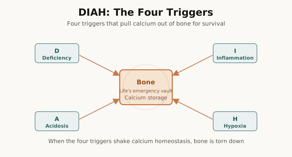
The essay that opens Part 2: Reversal: Why Calcium Makes Us Sick (The Same Calcium, the Opposite Result)
Introduction: What Changes the Direction of Calcium
In Part 1 we answered a single question. How does life handle calcium? Over 3.8 billion years life has built a sophisticated system for controlling calcium. The pumps of the cell membrane keep cytoplasmic calcium at 100 nanomoles, the storehouse called bone holds 99 percent of the body's total calcium, and parathyroid hormone, vitamin D, and calcitonin coordinate the flow of calcium throughout the body. At the synapse, calcium moves in and out on the scale of milliseconds and makes thought possible. In Chapter 1 we saw that the number 100 nanomoles is a fundamental design principle of life that has been conserved for 3.8 billion years; in Chapter 2 we saw that the Cambrian explosion was in effect a calcium revolution; in Chapter 3 we saw that, along with the advance onto land, a sophisticated hormone system evolved to manage the calcium bank called bone; and in Chapter 4 we saw that the refinement of calcium signaling became the material basis of the higher intelligence by which 86 billion neurons produce thought. Each stage was built upon the previous one, the ability to control calcium was itself the ability to survive, and it was a core element of evolutionary fitness.
In Part 2 we pose a new question. Why does this sophisticated system reverse? And how does that reversal connect to chronic disease? To answer this question, this book proposes the DIAH-7M pathway framework. It is a frame in which four triggers, Deficiency, Inflammation, Acidosis, and Hypoxia, cause calcium to flow out of the bones, and the leaked calcium leads to chronic disease through seven pathological mechanisms. Each of these components is a fact that science has already established. DIAH-7M is an integrated framework that fits these puzzle pieces into a single picture. Chapter 5 looks at the evolutionary background of this framework. Why did evolution choose calcium? What role does calcium play in the cycle of life? Chapter 6 deals in detail with the mechanism of each DIAH trigger, and Chapter 7 with each of the 7M pathological pathways.
Introduction: The Evolutionary Logic of Sacrificing Bone for the Sake of Survival
In Chapter 5 we looked at the big picture of the DIAH-7M pathway. DIAH is the English acronym for Deficiency, Inflammation, Acidosis, and Hypoxia. We proposed that these four states act as triggers that cause calcium to flow out of the bones. In this chapter we will look at the specific mechanism of each trigger and explore how calcium has served as an execution code for survival throughout the process of evolution.
How deficiency mobilizes bone calcium, how inflammation activates the bone-destroying cells, how acidosis dissolves bone, and how hypoxia changes bone metabolism. Each individual mechanism is already well studied scientifically. The contribution of this book is to connect them from a single integrated point of view.
Before we look at each trigger, we must first understand normal bone metabolism. To understand the abnormal, you must know the normal.
The Limits of Existing Medical Maps
Humanity has created great maps.
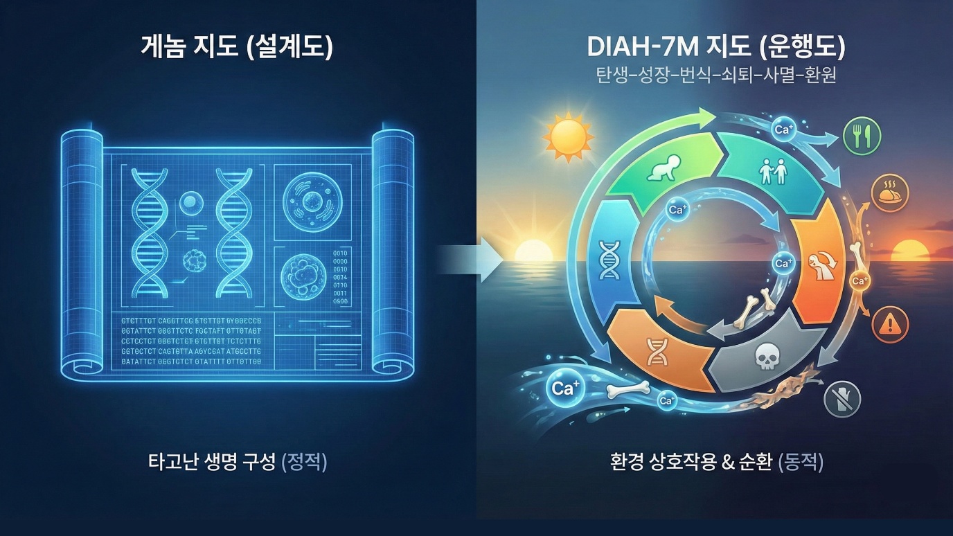
The genome map decoded all of the human genes. It read out 3 billion base sequences and revealed which gene performs which function and which mutation is associated with which disease. But the genome map focuses on "genes." It cannot explain why, even among identical twins who carry the same genes, one develops diabetes and the other does not.
The disease map (ICD, the International Classification of Diseases) systematically classified tens of thousands of diseases. It became possible for doctors around the world to diagnose and record disease in the same language. But the disease map focuses on "symptoms." High blood pressure is hypertension, high blood sugar is diabetes, an aching joint is arthritis. It puts a name on the symptom that has surfaced and prescribes a drug that suppresses that symptom.
Yet it cannot answer the most important questions. "How did this disease begin?" "Why, of all people, did this disease happen to me?" "Why do hypertension and diabetes come together?"
If you ask a doctor, the answer that comes back is mostly this. "Genetic factors, lifestyle, aging, and stress act in combination." It is a correct statement. But it is too abstract. There was no map that clearly explained through exactly which pathway a disease is made, why different diseases appear together, and that process.
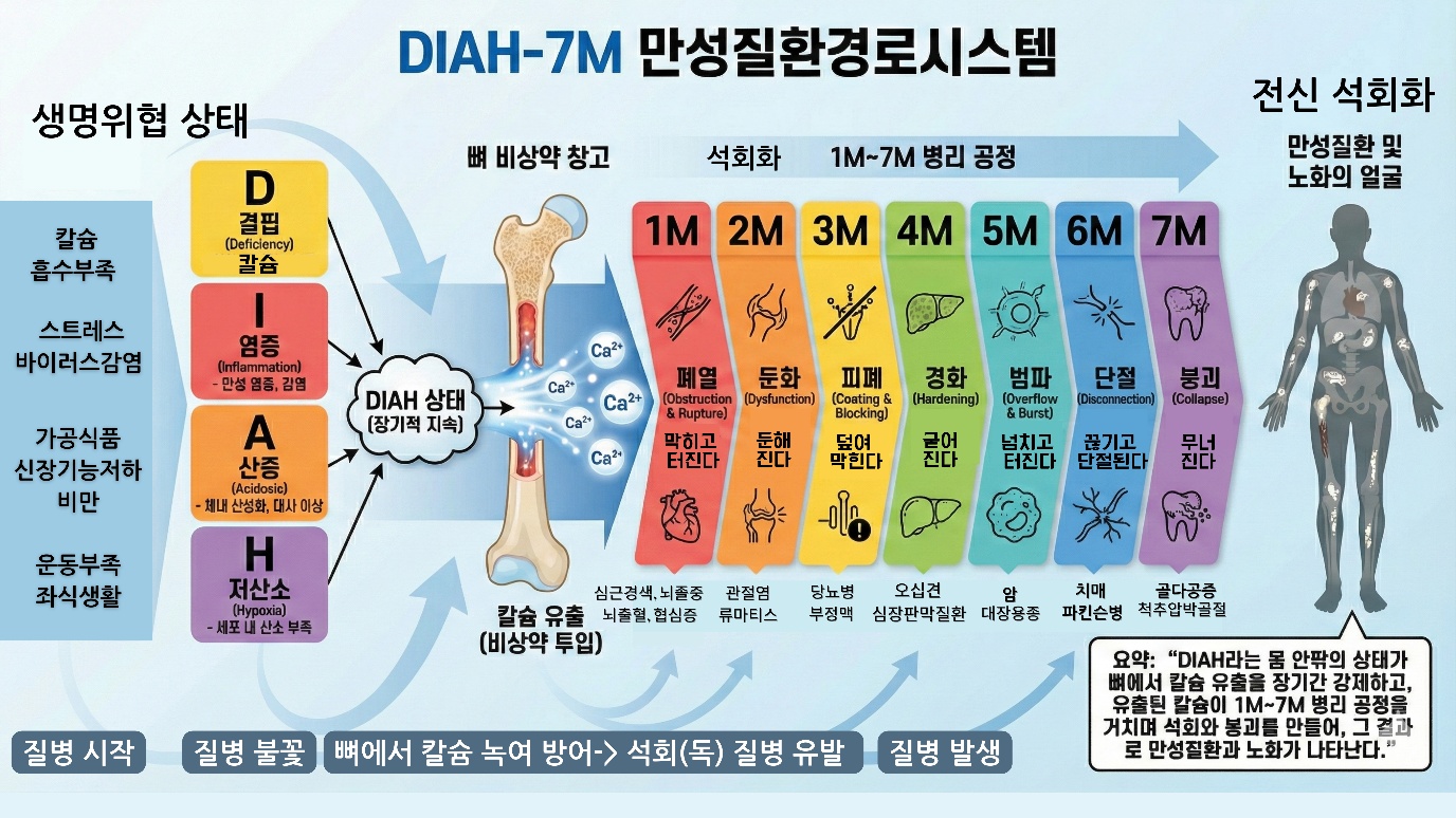
The DIAH-7M Aging and Chronic Disease Pathway System map presented in this chapter fills exactly that empty place and proves that calcium is the execution code of evolution. It shows not an abstract "cause" but a concrete "pathway." It does not classify symptoms but tracks a process.
The Basic Principles of Bone Metabolism
When we think of bone, a hard, unchanging material comes to mind. Like a dinosaur bone on display in a museum or a skeletal model in an anatomy lab, it seems like a material that will stay in place forever. But bone inside a living body is entirely different. Bone is a living tissue that changes ceaselessly.
About 10 percent of an adult's bone is replaced each year. Old bone is broken down and new bone is formed. This is called bone remodeling. Over a lifetime the entire skeleton is replaced several times. Bone is not a static structure but a dynamic tissue. Why is bone replaced ceaselessly? There are several reasons. The first is repair of damage. In everyday activity, microdamage accumulates in bone. Running, jumping, and even just walking create tiny cracks in bone. If this microdamage is left alone, it accumulates and eventually leads to fracture. Bone remodeling removes the damaged area and replaces it with new bone, maintaining structural integrity.
The second is mechanical adaptation. Bone adjusts its structure according to the load it receives. This is called Wolff's law. A region that receives a lot of load grows thicker, and a region that receives little load grows thinner. This is why an athlete's bones have higher density than an ordinary person's, and why an astronaut loses bone in a weightless environment. The third is calcium homeostasis. Bone is a calcium storehouse. When blood calcium is insufficient, it is drawn out of bone, and when there is a surplus, it is stored in bone. Bone remodeling makes this storage and release possible.
There are two main types of cell that carry out bone remodeling. Osteoclasts and osteoblasts. Bone is like a building under remodeling. The osteoclast is the demolition crew that breaks down the old wall, and the osteoblast is the construction crew that lays the cement. The osteoclast is the cell that breaks down bone.
As the name suggests, it is the cell that breaks bone. Osteoclasts derive from the immune cell lineage. Several precursor cells fuse to become a multinucleated giant cell, a large cell reaching 100 micrometers in diameter. The osteoclast attaches to the bone surface to form a sealing zone, and secretes hydrochloric acid within this zone. The pH drops to about 4.5. The strong acid dissolves hydroxyapatite, the mineral component of bone. At the same time, it secretes protein-degrading enzymes to break down the collagen matrix. The bone dissolves, leaving a hollow pit. The osteoblast is the cell that forms bone. It is the cell that makes bone. Osteoblasts derive from mesenchymal stem cells and differentiate from the same ancestor as fat cells and cartilage cells.
Osteoblasts secrete collagen and other proteins at the bone surface to form the bone matrix, and then calcium and phosphate deposit and hydroxyapatite crystals form, so that the bone hardens. Some osteoblasts become buried within the bone matrix they made, and these become osteocytes. Osteocytes are connected like a net within the bone, sensing mechanical stimuli and sending signals, and they play an important role in regulating bone remodeling.
Bone remodeling proceeds on a regular cycle. One remodeling unit is called the basic multicellular unit. The remodeling cycle proceeds as follows. First, in the activation phase, osteocytes sense a mechanical stimulus or microdamage and send a signal to recruit osteoclast precursor cells. Next is the resorption phase, in which osteoclasts arrive at the bone surface, differentiate, and over about 2 to 3 weeks resorb the bone, forming a hollow pit.
Then, in the reversal phase, the osteoclasts die and cleanup cells tidy the surface of the pit, preparing it for the osteoblasts. In the formation phase, osteoblasts arrive to fill the pit and secrete new bone matrix while calcification proceeds, a process that takes about 4 to 6 months. Finally, in the resting phase, remodeling is completed and the bone surface returns to a resting state. The whole cycle is about 4 to 6 months. Resorption is fast at 2 to 3 weeks, and formation is slow at 4 to 6 months. This asymmetry is important. The heart of normal bone remodeling is balance. Once the demolition crew, the osteoclasts, has dissolved the bone, the construction crew, the osteoblasts, comes to that spot and fills exactly that much bone. The demolition crew sends a signal to the construction crew that the work here is done, so come and fill it, and this is called coupling. It means the two work as a pair. But when a DIAH trigger is pulled, this coupling breaks. The demolition crew madly smashes the wall, but the construction crew is exhausted and does not come, or comes late. In the end only holes are left in the bone. This is the reality of osteoporosis.
The differentiation and activity of osteoclasts is regulated by a signaling system called RANKL-RANK-OPG. This is the core regulatory axis of bone metabolism. RANKL is the key signaling substance that promotes osteoclast differentiation, and osteoblasts, osteocytes, and activated immune cells produce RANKL. RANK is a receptor on the surface of osteoclast precursor cells. When RANKL binds to RANK, osteoclast differentiation begins, and the precursor cells fuse to become multinucleated osteoclasts.
OPG is the decoy receptor for RANKL. Osteoblasts and osteocytes secrete OPG, and OPG clings to RANKL and interferes with RANKL binding to RANK. Osteoclast differentiation is suppressed. The RANKL/OPG ratio determines the direction of bone remodeling. When RANKL is high and OPG is low, osteoclast activity increases and bone resorption dominates. When RANKL is low and OPG is high, osteoclast activity decreases and bone formation dominates. In the end, the switch that decides how many osteoclasts will be made to work, and for how long, is the RANKL/OPG ratio. The four DIAH triggers we will examine later push this switch in the same direction, that is, toward osteoclast activation, through different pathways.
Many hormones and signaling substances regulate the RANKL/OPG ratio. Estrogen increases OPG and decreases RANKL, protecting bone. Parathyroid hormone increases RANKL and promotes bone resorption. Inflammatory signaling substances such as tumor necrosis factor alpha, interleukin-1, and interleukin-6 increase RANKL and promote bone resorption.
Blood calcium concentration is precisely maintained in a range of about 8.5 to 10.5 mg/dL. Straying outside this narrow range endangers life. If calcium is too low, muscle cramps and cardiac arrhythmia occur, and if it is too high, cardiac arrhythmia and altered consciousness occur. There are three main hormones responsible for this precise regulation. Parathyroid hormone is secreted by the parathyroid glands, and when blood calcium drops, its secretion increases within minutes. Parathyroid hormone has three actions. It mobilizes calcium from bone to increase osteoclast activity, it increases calcium reabsorption in the kidney to reduce the calcium lost in urine, and it promotes vitamin D activation in the kidney.
Here there is an interesting paradox. When parathyroid hormone is continuously kept high, it activates osteoclasts and dissolves bone, but when a very low dose intermittently stimulates, it instead stimulates osteoblasts and increases bone formation. In fact, teriparatide, one of the osteoporosis drugs, uses this principle by injecting a portion of parathyroid hormone once daily. The problem is that in a state of chronic deficiency, parathyroid hormone is continuously elevated. Active vitamin D increases calcium absorption in the intestine and increases the expression of calcium-binding proteins and calcium channels.
At high doses it also promotes the mobilization of calcium from bone. Calcitonin is secreted by the C cells of the thyroid, and when blood calcium rises it is secreted and directly suppresses osteoclast activity. However, the physiological role of calcitonin appears to be less important than that of parathyroid hormone or vitamin D. It is important to understand the priorities of this system. Maintaining blood calcium takes priority over maintaining bone.
When blood calcium drops, parathyroid hormone draws calcium out of the bone. Even if the bone weakens, it maintains blood calcium. This is because if blood calcium drops you can die immediately, whereas the weakening of bone is a later problem. This is the fundamental reason that calcium deficiency leads to bone loss.
The Redefinition of the Key Substances: The True Face of Calcium, Bone, and Calcification
The greatest originality of the DIAH-7M Aging and Chronic Disease Pathway System is that it looks at the three key substances we believe we already know, calcium, bone, and calcification, from an entirely different perspective and redefines them functionally. Without this redefinition, we cannot understand why and how chronic disease is made.
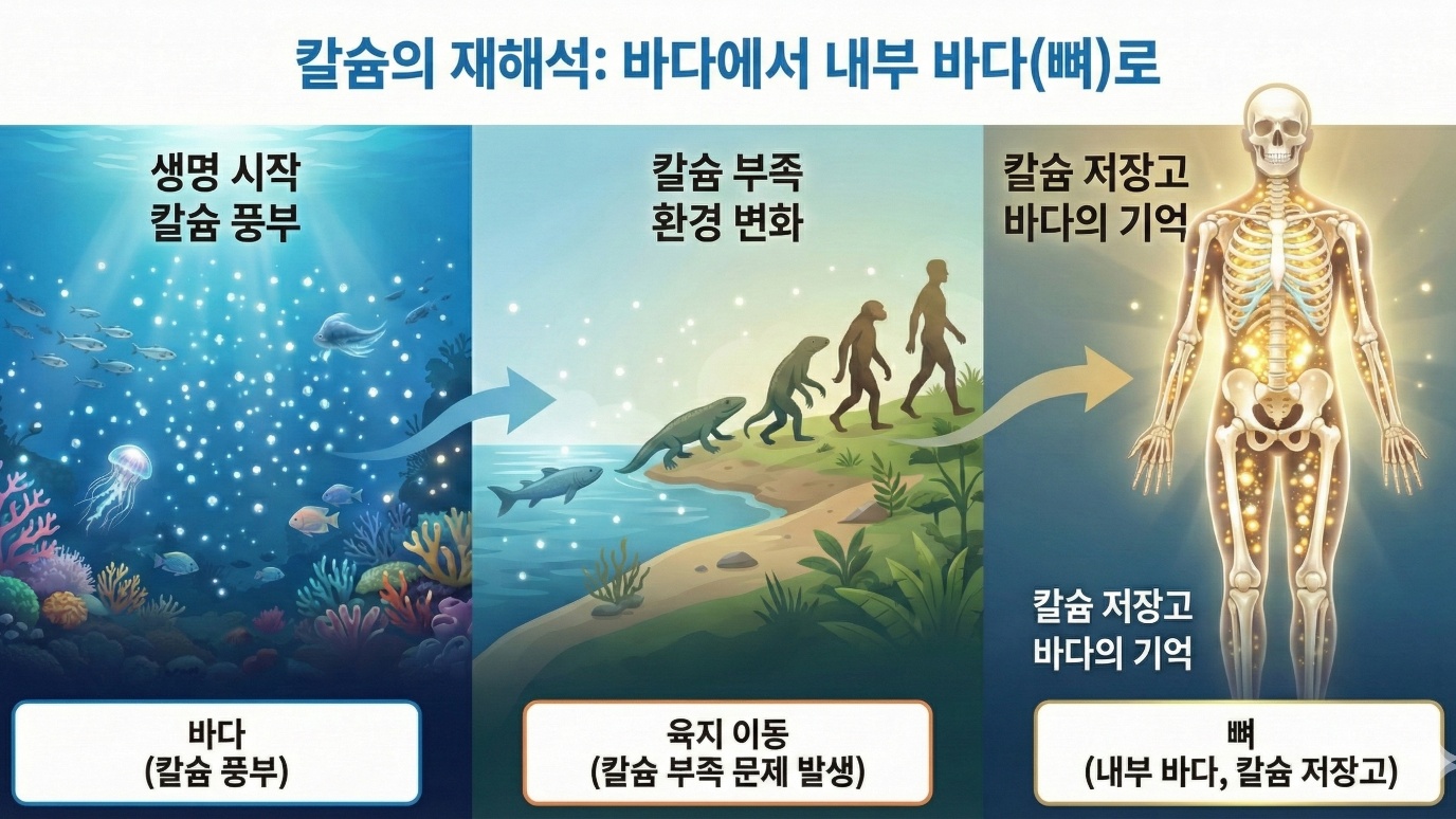
First, Calcium: Not a Mere Nutrient but the "Currency of Life"
Calcium has until now been perceived as a nutrient, a mineral of sorts, that we make a point of eating to strengthen our bones. But the true face of calcium is entirely different.
Calcium is the currency of life that makes the heart beat, moves the muscles, transmits nerve signals, and operates the immune cells. Just as the economy stops when there is no money, life activity stops when there is no calcium. When blood calcium concentration strays outside the normal range (8.5 to 10.5 mg/dL), cardiac arrhythmia occurs, and in severe cases cardiac arrest can come. That is how essential calcium is to survival itself.
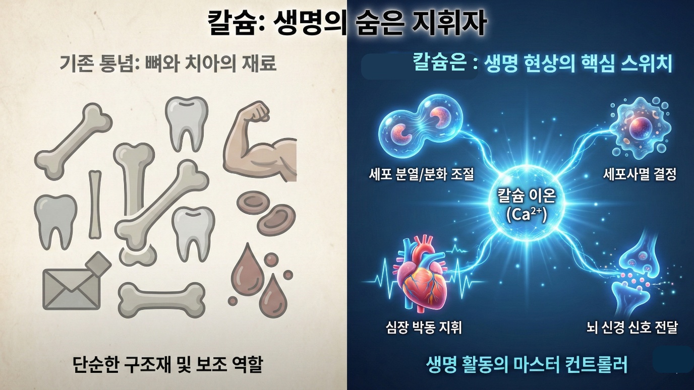
This is the reason we compare the flow of calcium to the "flow of money" in this chapter. Income is cut off, spending soars, the emergency fund is drawn down, unpaid debt piles up, and in the end it reaches bankruptcy. In our body too, the very same thing happens by way of calcium.
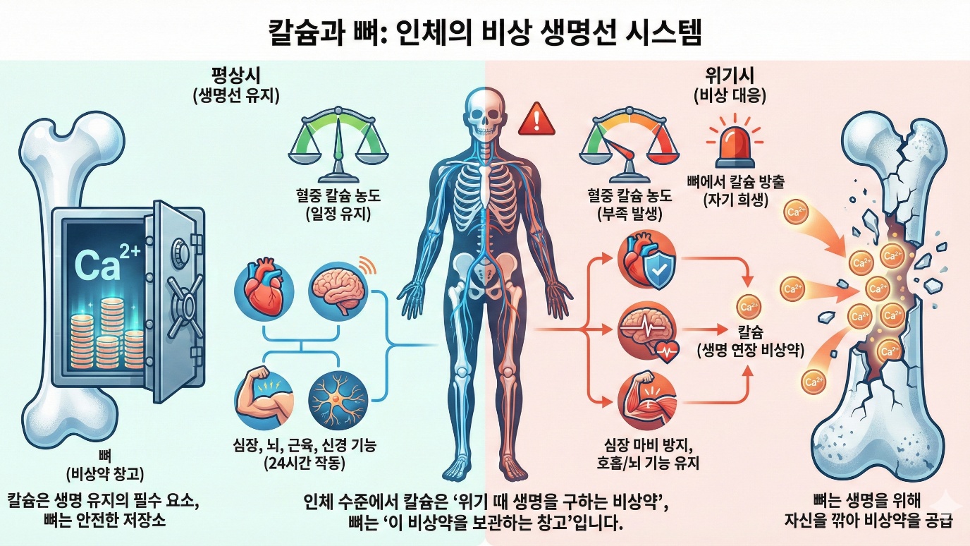
Second, Bone: Not a Support but the "Emergency Warehouse of Life"
In conventional wisdom, bone was regarded as the skeleton that holds the body upright, or a warehouse of sorts that stores calcium. But the true role of bone is far more decisive.
Bone is a giant vault in which 99% of the body's total calcium is stored. Only 0.5g of calcium circulates in the blood, but about 1kg of calcium, give or take, is stockpiled in the bones. Bone is also the pillar that supports the body, but more essentially it is the bank that keeps the emergency fund of life.
At every moment of DIAH crisis, when blood calcium concentration drops, inflammation spreads, body fluids tilt toward acidity, and cells fall into a hypoxic state, the bone shaves off a little of itself and sends out calcium. Therefore osteoporosis is not a mere phenomenon of aging. It is a withdrawal record that shows how fiercely, over a long time, my body has opened the door of this warehouse and waged a struggle for survival.
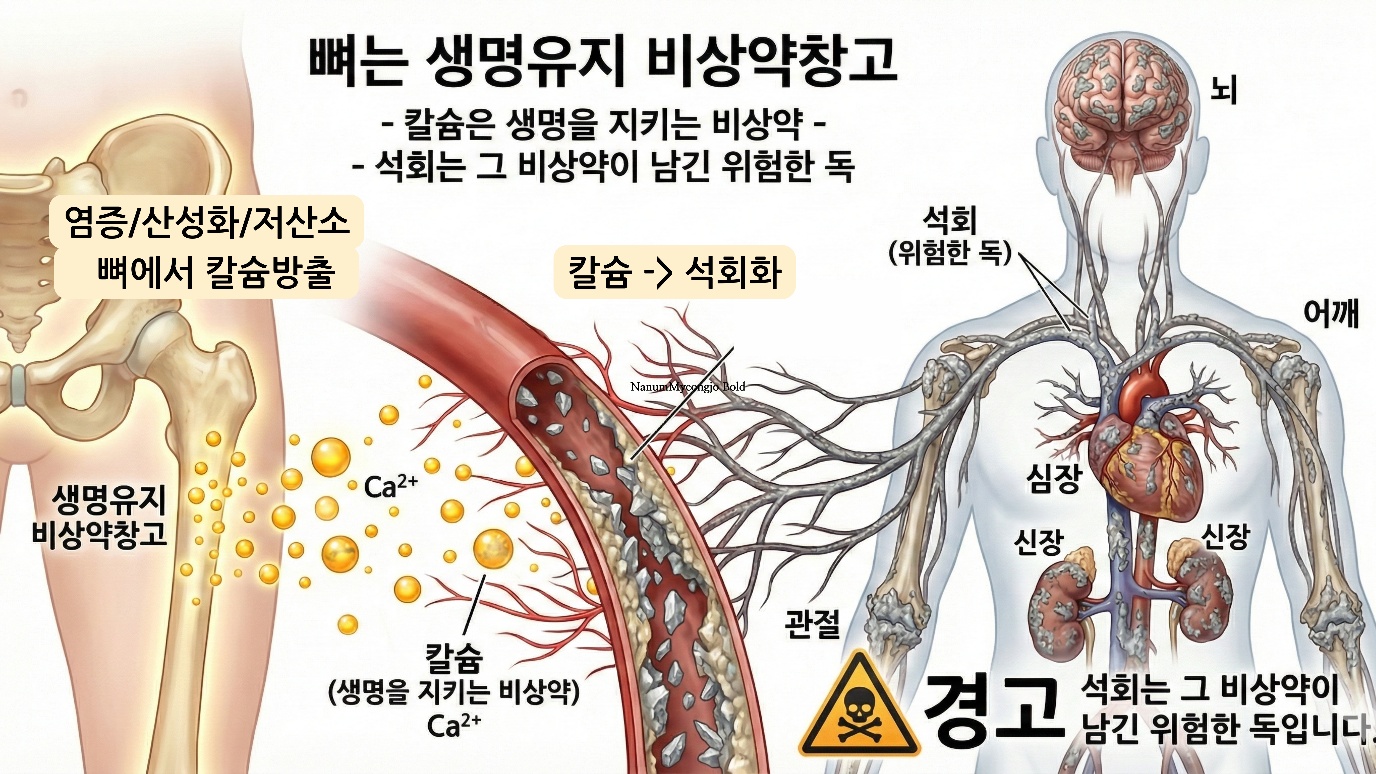
Third, Calcification: Not a Residue but the "Raw Material of the Pathological Mechanism" and a "Coordinate"
In the existing view, the calcification and stones that form throughout the body, in blood vessels, joints, and organs, were regarded as the stones, residue, and byproducts of sorts that inevitably form as we age. They were treated not as the center of disease but as an incidental phenomenon. This conventional wisdom must be overturned. When it leaves the bone, calcium is an emergency medicine that saves life. But the calcium that remains after finishing its role, unable to return to its place, becomes poison and piles up as calcification. In blood vessels that should be soft, in joints that should be flexible, in organs that should be tender, a lump of calcium as hard as stone deposits.
This calcification is precisely the raw material of chronic disease. At the same time it is the coordinate of pathology and a warning signal that tells us at which part of the body, and how far, collapse has progressed.
That calcification is visible somewhere in my body does not simply mean that one lump of stone has formed. It is a living record that shows how long, and how often, that spot has been attacked by calcium, the bone's emergency medicine, within the DIAH environment.
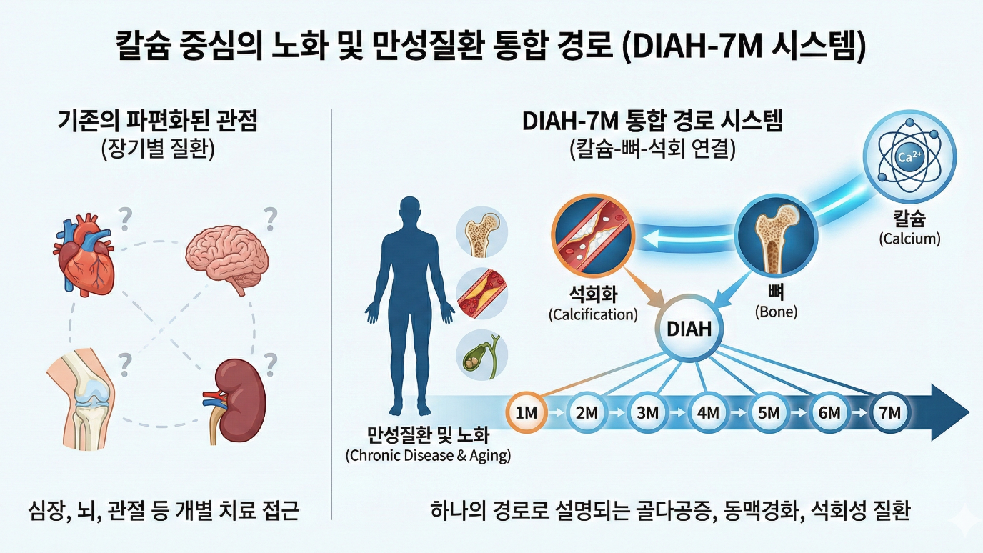
DIAH-7M: A New Map That Reads Disease Through the Flow of Calcium
Now we have confirmed the true face of the three key substances. DIAH-7M is the very system that explains the entire process of chronic disease through this flow of calcium.
• Why does calcium flow out of the bones? (the DIAH triggers)
• Where does the leaked calcium go? (efflux and overflow)
• Where does it pile up? (calcification deposits)
• How does it become disease? (the 7M mechanisms)
If existing maps showed "what is there," DIAH-7M shows "how it progresses." From the initiating factor of disease to the final onset, it tracks the whole process in seven stages, following the single substance called calcium.
It explains, in the language of calcium, why hypertension, diabetes, and dementia come together, their common root. This is the original distinguishing point of DIAH-7M.
DIAH-7M: A Closer Look
| Abbreviation | English | Korean | Action | Description |
|---|---|---|---|---|
| D | Deficiency | 결핍 | Bone calcium fills the shortfall | Deficiency of calcium, minerals, vitamin D, and the like → replenished by breaking down calcium from bone → blood → supplied to various cells ※ Common: D, I, A, H all cause a drop in blood calcium, which is replenished by breaking down calcium from bone. |
| I | Inflammation | 염증 | Calcium is mobilized to put out inflammation | Massive consumption of calcium in the inflammatory response → replenished by breaking down calcium from bone → blood → concentrated at the site of inflammation |
| A | Acidosis | 산증 | Bone is sacrificed to neutralize acid | Alkaline calcium used to neutralize acid → replenished by breaking down calcium from bone → blood → whole-body pH balance |
| H | Hypoxia | 저산소 | With oxygen shortage, calcium floods excessively into the cell | Cellular oxygen shortage → calcium pump failure → blood calcium over-absorbed into the cell → blood calcium deficiency → replenished by breaking down calcium from bone → blood → re-absorbed by damaged cells → vicious cycle |