
This piece is the front part of Chapter 11 of Stones in the Body: More Frightening Than Cancer (Yoon Jong-won). It is a narrative containing the author's academic hypothesis, and the body, figures, and citations follow the manuscript as written.
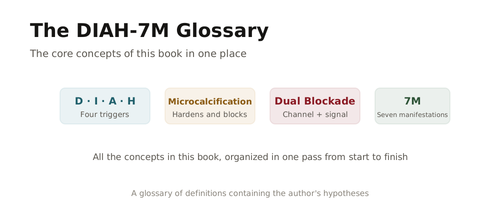
An integrated pathophysiology model in which calcium efflux from bone is triggered, and the leaked calcium, through seven pathological mechanisms, deposits and accumulates in soft tissue, taking part in the onset and worsening of chronic disease
1. DIAH Triggers (four causes of calcium efflux)
The four triggers D·I·A·H all induce a drop in blood calcium,
and the shortfall is replenished by breaking down calcium from bone.
| Abbreviation | English | Korean | Action | Description |
|---|---|---|---|---|
| D | Deficiency | Deficiency | Bone calcium fills the shortfall | Deficiency of calcium and minerals, vitamin D, and so on → replenished by breaking down calcium from bone → blood → supplied to various cells ※ Common note: D (Deficiency) is the representative path that can directly create a 'drop' in blood calcium, while I (Inflammation)·A (Acidosis)·H (Hypoxia), depending on the situation, shake the 'consumption·distribution·ionization (availability)' of calcium and as a result trigger or accelerate the D path (PTH-bone mobilization) into operating more often |
| I | Inflammation | Inflammation | Calcium is mobilized to quell inflammation | Massive consumption of calcium in the inflammatory response → replenished by breaking down calcium from bone → blood → concentrated at the site of inflammation |
| A | Acidosis | Acidosis·Oxidation | Bone is sacrificed to neutralize acid | 1) Neutralizing acidity (pH↓): alkaline calcium is used to neutralize acid → replenished by breaking down calcium from bone → blood → whole-body pH balance 2) Neutralizing oxidation (ROS): oxidative stress → massive consumption of calcium in the inflammatory response → replenished by breaking down calcium from bone → blood → concentrated at the site of inflammation |
| H | Hypoxia | Hypoxia | Under oxygen shortage, calcium flows excessively into the cell | Cellular oxygen shortage → decline of Ca²⁺ pump/regulatory function due to reduced ATP → intracellular calcium overload (damage·death signal) → during tissue damage and recovery, calcium demand·distribution is shaken → as a result, when blood calcium 'availability' becomes unstable, the D path (PTH-bone mobilization) is easily summoned → if repeated, a vicious cycle |
The four triggers D·I·A·H organized in this table look, on the surface, like different causes, but in fact they all work in one common direction, that of "a force that lowers the blood calcium concentration," and their role is to forcibly flip the switch so that calcium is drawn out of the bone to make up for the calcium that has fallen.
According to several endocrinology textbooks and international bone-metabolism societies, the heartbeat and the electrical signals of nerve and muscle are designed to be so sensitive that a disturbance arises the instant the blood calcium concentration wavers even slightly, and so, when our body enters a deficiency (D) state in which it cannot receive enough calcium, vitamin D, and minerals from food, it immediately sends a signal to the bone storehouse through parathyroid hormone (PTH), ordering it to "make an emergency withdrawal of calcium to fill the shortfall," and in an inflammation (I) situation where chronic inflammation is progressing, because calcium is consumed in large amounts to maintain the immune response, the calcium in the blood is quickly used up, and the system moves in the direction of again drawing that shortfall from the bone and concentrating it around the site of inflammation.
Also, in the acidosis (A) stage, where acidic waste accumulates in the body through an acidic diet·overwork·metabolic disorder and so on, according to several physiology sources, because the calcium inside the bone acts as a powerful alkaline buffer and is mobilized to neutralize the acid in the blood, even though on the surface it is felt as no more than excess stomach acid·reflux·fatigue, internally, under the name of "a sacrifice to catch the acid," calcium is being cut away from the bone bit by bit, and in the hypoxia (H) situation, where microvessels are blocked and tissue cannot breathe properly, as the calcium pump inside the cell and the energy system that maintains the membrane potential break down, the movement of calcium between blood and cell loses its control and leaks out, and in the end, to make up for the blood calcium shortage, the breakdown of bone calcium is once again accelerated, as is known.
Seen this way, the DIAH triggers are not four causes standing apart from one another but can be called "the last safety device the body chooses when I have failed to eat, or inflammation has flared up, or acid has accumulated, or tissue can no longer breathe," and because that safety device is always released in the direction of opening the bone and drawing out calcium to use, the core message the DIAH trigger table conveys is that today's stress and diet, chronic inflammation and breathless living all combine and in the end lead into a single road that slowly melts the bone away.
When these four switches turn on and off repeatedly, the calcium that comes out of the bone leads directly into the 7M mechanisms, creating the pathway of chronic disease that blocks blood vessels, hardens tissue, and brings down organs and nerves, and in what follows we will trace, step by step, how this DIAH and 7M connect and reveal themselves as actual disease in our body.
[ The DIAH Trigger Sub-classification System ]
Each DIAH trigger is subdivided into sub-types as below, and through this the specific cause of a chronic disease can be traced.
■ D (Deficiency) Sub-classification
| Code | Name | Core Mechanism |
|---|---|---|
| D-PH | Physical Stimulus Deficiency | Lack of gravity·weight-bearing·muscle-contraction stimulus → the bone-stimulus signal is cut off - sedentary/lack of standing·walking (weight-bearing time↓), lack of strength exercise (muscle-contraction load↓), bed rest·long-term hospitalization·cast/immobilization, sarcopenia·muscle-strength decline, weightlessness·no-load, avoidance of activity due to chronic pain·joint disease, reduced walking/loading due to nervous-system problems (Parkinson's·stroke aftereffects), and so on |
| D-NT | Nutrient Deficiency | Shortage of calcium·vitamin D·K·Mg·protein and so on - shortage of calcium·vitamin D·K·Mg, protein shortage, reduced absorption (aging-related decline in gastric acid secretion·intestinal disease·gastrectomy), excess sodium·caffeine (excretion↑), low-weight/sarcopenic-type nutritional deficiency (intake itself↓), heavy drinking (D-NT/A-DT: nutrient absorption↓ + acid load↑), and so on |
| D-LF | Lifestyle Input Deficiency | Shortage of rhythm·recovery such as sleep·sunlight·hydration - sleep shortage/quality decline, lack of sunlight exposure, hydration shortage, chronic stress (D-LF/I-ST - recovery↓+inflammation↑), recovery time↓, and so on |
| D-MD | Medication-induced Deficiency | Absorption↓/excretion↑/metabolic disturbance caused by steroids·PPIs·diuretics and so on - steroids, PPIs (gastric-acid suppressants), diuretics·anticonvulsants, thyroid hormone excess (including over-supplementation), and so on |
| D-HR | Hormone Deficiency | Deficiency of estrogen·testosterone·growth hormone → weakening of the osteoclast brake - menopause, decline of male hormone, decrease of growth hormone with aging, and so on |
D-PH (Physical Stimulus Deficiency) occurs when gravity and weight-bearing load are not applied to the bone due to lack of exercise·sedentary living·long-term hospitalization·weightless environment and so on. When bone receives pressure, it generates a micro-current through the piezoelectric effect (Piezoelectricity), and this signal activates osteoblasts to maintain·strengthen the bone. The phenomenon in which an astronaut's bone density decreases by 10 to 20% after a six-month stay is representative evidence of D-P.
※ D-HR (Hormone Deficiency): Deficiency of estrogen·testosterone·growth hormone directly affects bone metabolism. The decrease of estrogen after menopause, the decline of testosterone in men, and the decrease of growth hormone with aging are all equivalent to a weakening of the brake that suppresses osteoclast activity. Together with D-NT (Nutrient Deficiency), this is a major pathway of bone-calcium efflux.
※ Interpretation of PTH excess: An excess of parathyroid hormone (PTH) is not an independent trigger but a compensatory response to D (Deficiency). When blood calcium falls, PTH is secreted and draws calcium out of the bone, which is a mechanism that appears not as a cause but as a result of D.
■ I (Inflammation) Sub-classification
| Code | Name | Core Mechanism |
|---|---|---|
| I-EN | Environmental Amplifier | Increase of inflammatory burden from pollution·chemicals·dust·electromagnetic waves (under research) and so on - fine dust·air pollution, chemicals·environmental hormones, occupational exposure (solvents/dust/metals), electromagnetic-wave/high-frequency exposure (E), and so on |
| I-MT | Metabolic Inflammation | Inflammatory substances or metabolic waste generated inside the body such as obesity·glucotoxicity·insulin resistance - obesity·visceral fat, insulin resistance, glucotoxicity (AGEs), ultra-processed-food-centered eating habits, dyslipidemia·fatty liver, and so on |
| I-CH | Chronic Infection | Persistent infection such as periodontitis·leaky gut·chronic bronchitis - periodontitis·sinusitis (oral cavity/upper airway), leaky gut·gut-flora imbalance (gut), chronic bronchitis (lower airway), urinary-tract infection/chronic cystitis (urinary tract), and so on |
| I-AG | Age-related Inflammation | Chronic inflammation due to immune aging) - underlying: immune aging (inflammaging), accumulation of cellular waste, decline of antioxidant capacity, sarcopenia·reduced activity (lack of exercise ↔ aging-inflammation vicious cycle), and so on |
| I-ST | Stress-induced Inflammation | Inflammation due to collapse of the cortisol rhythm · chronic stress (D-LF/I-ST - recovery↓+inflammation↑), sleep shortage·apnea (inflammation↑+acidosis↑+hypoxia↑), autonomic-nervous imbalance, and so on |
| I-AI | Autoimmune Inflammation | Chronic inflammation due to an autoimmune response - rheumatoid arthritis, lupus (SLE), ankylosing spondylitis, psoriasis, inflammatory bowel disease (Crohn's disease, ulcerative colitis), multiple sclerosis, Sjögren's syndrome, and so on |
※ The link between hormone excess and inflammation: Cortisol excess (chronic stress) suppresses inflammation in the short term, but when it becomes prolonged it results in a failure of inflammation control (I-ST). Also, glucocorticoid drugs (steroids) suppress osteoblasts and prolong the lifespan of osteoclasts, inducing D-MD and I-ST at the same time. Therefore "hormone excess" is interpreted not as an independent trigger but as an upstream cause that induces I.
※ I-AI (Autoimmune Inflammation): In autoimmune disease, the immune system attacks the body's own tissue and induces chronic inflammation. In rheumatoid arthritis, lupus, ankylosing spondylitis, and so on, inflammatory cytokines (TNF-α, IL-1, IL-6) activate osteoclasts and promote bone-calcium efflux. Unlike I-CH (Chronic Infection), this is inflammation caused by an internal immune abnormality without an external pathogen.
■ A (Acidosis) Sub-classification
| Code | Name | Core Mechanism |
|---|---|---|
| A-FN | Functional Acidosis | Insufficient acid-excretion capacity due to decline of kidney·liver function - chronic kidney disease, decline of liver function, dehydration·diarrhea, poorly controlled diabetes (ketone/acid-load risk), and so on |
| A-DT | Dietary Acid Load | Increase of the body's acid load from high-protein·high-salt·processed food and so on - high-protein·low-vegetable (when accompanied by insufficient alkaline input), ultra-processed·high-salt·high-sugar, heavy alcohol drinking (nutrient↓+acid↑), and so on |
| A-RS | Respiratory Acidosis | Imbalance caused by CO₂ stagnation (COPD, sleep apnea) - COPD, sleep apnea (inflammation↑+acid↑+oxygen↓), respiratory disease (carbon-dioxide accumulation), and so on |
| A-HM | Hypermetabolic Acidosis | Hyperthyroidism → metabolic hyperactivity → increase of acid production - rise of basal metabolic rate → oxygen consumption↑ → metabolic products (acid)↑ → mobilization of alkali (calcium) from bone |
※ A-HM (Hypermetabolic Acidosis): Hyperthyroidism excessively promotes metabolism and increases acid production. Through the path of rise of basal metabolic rate → increase of oxygen consumption → increase of metabolic products (acid) → mobilization of alkali (calcium) from bone, it contributes to bone-calcium efflux via the A trigger.
■ H (Hypoxia) Sub-classification
| Code | Name | Core Mechanism |
|---|---|---|
| H-CR | Circulatory Hypoxia | Circulatory function of heart·blood vessels·muscle↓ → blood does not circulate - heart failure, vascular disease, lack of exercise (→blood-flow stagnation), thrombus, dehydration/bleeding, and so on |
| H-RS | Respiratory Hypoxia | Oxygen-supply capacity of lungs·blood↓ - lung disease, anemia (oxygen transport↓), sleep apnea (inflammation↑+acid↑+oxygen↓), smoking·carbon monoxide, and so on |
| H-AN | Anemia-induced Hypoxia | Decline of the blood's oxygen-transport capacity - iron-deficiency anemia, anemia of chronic disease, vitamin-B12/folate-deficiency anemia, hemolytic anemia, chronic-kidney-disease anemia (EPO↓), and so on |
H (Hypoxia) is a low-perfusion·low-oxygen microenvironment, the final trigger point that ushers entry into the 7M mechanisms and at the same time an entry gate that amplifies I (Inflammation) through feedback. In particular, lack of exercise delivers a double blow, cutting off the bone signal through D-PH (Physical Stimulus Deficiency) while at the same time stopping the whole body's oxygen delivery through H-CR (Circulatory Hypoxia).
※ H-AN (Anemia-induced Hypoxia): Anemia lowers the blood's oxygen-transport capacity and induces tissue hypoxia. Iron-deficiency anemia, anemia of chronic disease, vitamin-B12/folate-deficiency anemia, chronic-kidney-disease anemia (EPO decrease), and so on fall under it. Unlike H-CR (Circulatory type: blocked blood vessels) and H-RS (Respiratory type: decline of lung function), it is an oxygen-transport problem of 'the blood itself.' The chain path of D-NT (Nutrient Deficiency) → iron/B12 deficiency → H-AN (Anemia-induced Hypoxia) is common.
※ DIAH interpretation of hormonal imbalance: "Hormone excess/deficiency" is not an independent fifth trigger. Hormone deficiency (estrogen, testosterone, growth hormone) is included in D, and PTH excess is classified as a compensatory response (mechanism) to D. Cortisol excess (chronic stress) is an upstream cause that induces I, and hyperthyroidism is interpreted as the metabolic hyperactivity that induces A. In this way, every hormone-related pathology can be explained within the DIAH system.
※ This coding system is to be continuously validated and upgraded through future clinical research.
※ The primary/secondary/tertiary classification and coding system (19 codes): a new proposal by the DIAH-7M Laboratory (clinical validation needed)
※ Trigger ↔ factor relationship: based on existing medical research (PMC/paper validation complete)
※ Explanation of the 7M term:
• 폐열 (閉裂, Obstruction and Rupture) is a term different from the 폐열 (肺熱, lung heat) of Korean medicine; it is a coined word meaning 'obstruction (閉)' and 'rupture (裂).' It encompasses luminal obstruction (blocking of blood vessels/airway/intestinal tract), rupture (aneurysm/organ bursting), and hemorrhage (internal bleeding/external bleeding).
■ Rules for Using the Other (OT) Code
| No | Content |
|---|---|
| 1 | If it does not fall under an existing code by 50% or more, use OT |
| 2 | When using OT, a note of the specific factor is required (e.g., I-OT autoimmune) |
| 3 | When the same factor accumulates 5 or more OT entries → review for a new code |
| 4 | Review reclassification of OT items once a year |
※ The OT (Other) code is a 'temporary holding box' for factors that do not clearly fit the existing classification. It is a management principle that prevents indiscriminate use and, once accumulation exceeds a certain standard, promotes them into a new formal code.
■ Code Notation and Compound-coding Principles
| Item | Content |
|---|---|
| Format | [Trigger]-[Sub-code]-[Order] (e.g., I-MT-2 = Metabolic Inflammation, secondary) |
| Compound example | I-MT-2 (main) + D-PH-2 (sub) + A-RS-1 (sub) |
| Main code | Leading trigger = the trigger that operated first in time |
| Sub-code | Accompanying trigger = a trigger activated afterward (up to 3 recommended) |
※ Used when multiple triggers are involved in a single disease. The trigger that operated first in time is marked as the 'main code,' and the trigger that chained afterward as the 'sub-code.' Example: a case that started with stress (I-ST) and was worsened by lack of exercise (D-PH) ※ I-ST-1 (main) + D-PH-2 (sub)
■ Order-determination Criteria
| Order | Determination Question | Applicable Examples |
|---|---|---|
| Primary | Is a mind/stress/rhythm problem the root cause? | chronic stress, depression, anxiety, sleep-rhythm collapse, trauma |
| Secondary | Is it the result of lifestyle habits accumulated daily? | lack of exercise, overeating, unbalanced diet, smoking, drinking, sedentary living |
| Tertiary | Is an acute event/trauma/medical intervention the cause? | traffic accident, fall, surgery, acute infection, drug side effect |
※ Even for the same trigger, the order changes depending on 'where it started.' If a mind problem is the root, it is primary; if lifestyle accumulation, secondary; if a sudden event, tertiary. Order determination becomes an important clue for establishing a treatment strategy.
■ DIAH Trigger Code Determination Procedure (6 steps)
| Step | Question and Determination | Code (English) | Name (Korean) |
|---|---|---|---|
| Step 1 | Confirm the main symptom/disease: what is the patient's chief complaint and diagnosis? | - | e.g., hypertension, diabetes, osteoporosis, depression |
| Step 2 | Determine the leading DIAH trigger: which of D, I, A, H operated first? | D, I, A, H | Deficiency, Inflammation, Acidosis, Hypoxia |
| Step 3 | Trace the trigger cause: what is the specific cause of that trigger? | - | e.g., lack of exercise, eating habits, stress, infection |
| Step 4 | Determine the order: which level does the cause fall into? | 1st, 2nd, 3rd | Primary (root), Secondary (habit), Tertiary (acute) |
| Step 5 | Select the sub-code: choose the code matching trigger + cause type | D-PH, I-MT, A-DT, H-CR, etc. | Physical stimulus, Metabolic, Dietary, Circulatory, etc., 19 in total |
| Step 6 | Compound coding: when multiple triggers exist, mark main code + sub-code | e.g., I-MT-2 + D-PH-2 | Metabolic inflammation (main) + Physical-stimulus deficiency (sub) |
※ This is the standard procedure that starts from the patient's symptoms and assigns a code step by step. It traces the cause in the flow of 'what hurts ※ why it hurts ※ since when it has hurt.'
■ Trigger × Order Matrix (●=main, ○=possibly applicable)
| Trigger | Code (English) | Name (English/Korean) | Primary | Secondary | Tertiary |
|---|---|---|---|---|---|
| D | D-PH | Physical Stimulus Deficiency | ● | ○ | |
| D-NT | Nutrient Deficiency | ● | ○ | ||
| D-LF | Lifestyle Input Deficiency | ● | ● | ||
| D-MD | Medication-induced Deficiency | ○ | ● | ||
| D-OT | Other Deficiency | ○ | ○ | ○ | |
| I | I-EN | Environmental Amplifier | ● | ○ | |
| I-MT | Metabolic Inflammation | ● | |||
| I-CH | Chronic Infection | ● | |||
| I-AC | Acute Infection | ● | |||
| I-AG | Age-related Inflammation | ● | |||
| I-ST | Stress-induced Inflammation | ● | |||
| I-OT | Other Inflammation | ○ | ○ | ○ | |
| A | A-FN | Functional Acidosis | ○ | ● | |
| A-DT | Dietary Acid Load | ● | |||
| A-RS | Respiratory Acidosis | ○ | ● | ○ | |
| A-OT | Other Acidosis | ○ | ○ | ○ | |
| H | H-CR | Circulatory Hypoxia | ● | ○ | |
| H-RS | Respiratory Hypoxia | ○ | ● | ○ | |
| H-OT | Other Hypoxia | ○ | ○ | ○ |
※ It shows at a glance in which order the 19 sub-codes mainly operate. ● is the representative order of that code, and ○ is an order that may apply depending on the situation. Example: I-ST (Stress-induced Inflammation) mainly starts at the primary (root) order.
■ Explanation of the New Codes
| Code | Name | Description |
|---|---|---|
| I-AC | Acute Infection | Inflammation due to acute infection such as pneumonia·sepsis·acute inflammation. Falls under the tertiary (acute) trigger |
| D-OT, I-OT A-OT, H-OT | Other | Unclassified factors that do not fall under an existing code by 50% or more. For securing classification completeness (MECE) |
※ This is an explanation of the 5 codes (I-AC, D-OT, I-OT, A-OT, H-OT) added to the existing 14 codes. I-AC is a code for acute infection, and the OT codes are 'other' codes for unclassified factors.
■ Final Code Classification Table (19 codes)
| Trigger | Number of Codes | Code List |
|---|---|---|
| D (Deficiency) | 5 | D-PH (Physical stimulus), D-NT (Nutrient), D-LF (Lifestyle input), D-MD (Medication-induced), D-OT (Other) |
| I (Inflammation) | 7 | I-EN (Environmental), I-MT (Metabolic), I-CH (Chronic infection), I-AC (Acute infection), I-AG (Aging), I-ST (Stress-induced), I-OT (Other) |
| A (Acidosis) | 4 | A-FN (Functional), A-DT (Dietary), A-RS (Respiratory), A-OT (Other) |
| H (Hypoxia) | 3 | H-CR (Circulatory), H-RS (Respiratory), H-OT (Other) |
※ This is the complete list of the total 19 sub-codes under the 4 DIAH triggers. It consists of D (Deficiency) 5, I (Inflammation) 7, A (Acidosis) 4, and H (Hypoxia) 3.