
Continuing from the previous piece, we look at the point where the DIAH triggers and the 7M pathological mechanisms connect into a single common pathway. The references cited follow the manuscript as written.
The DIAH-7M Pathway: The Common Pathway of Aging and Chronic Disease
Modern medicine classifies diseases individually. Cardiovascular disease is handled by cardiology, osteoporosis by endocrinology or orthopedics, arthritis by rheumatology, kidney failure by nephrology, and dementia by neurology. Each is diagnosed and treated in a different way by a different specialist. This specialization has made deep understanding and expert treatment of each disease possible. But when we step back and look at the whole picture, an interesting pattern appears.
Many of these diseases share a common element. It is calcification. Arterial calcification is related to cardiovascular disease, joint calcification to arthritis, kidney calcification to kidney failure, and brain calcification to cognitive decline. Why are so many chronic diseases connected to calcification? Is it coincidence, or is there a common pathway? The DIAH-7M pathway framework answers this question by connecting the scientific facts that have already been established.
| Abbreviation | English | Korean | Action | Explanation |
|---|---|---|---|---|
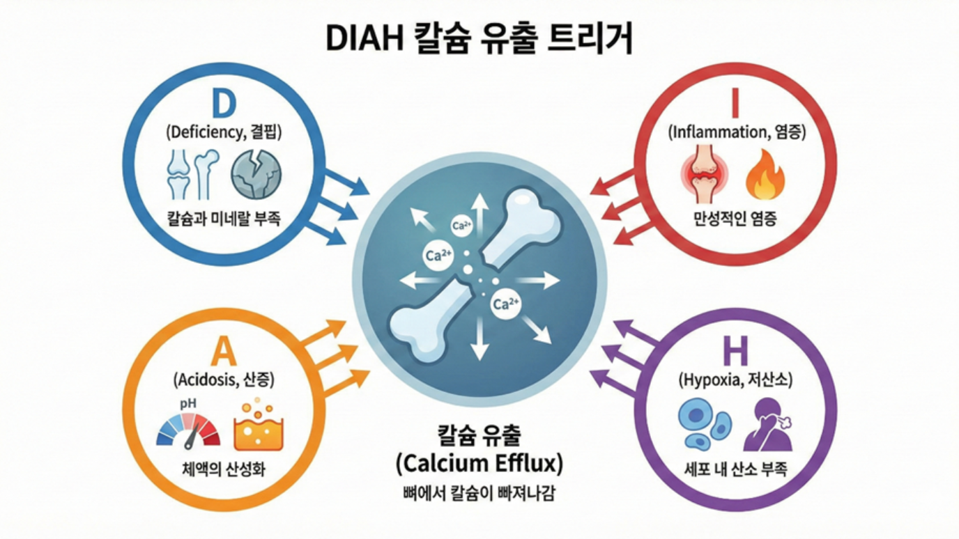
| D | Deficiency | 결핍 | Bone calcium fills the shortfall | Deficiency of calcium, minerals, vitamin D, and the like leads to calcium being broken down from the bones to replenish it, then supplied through the blood to the various cells. * Common to all: D, I, A, and H all induce a drop in blood calcium, which is replenished by breaking down calcium from the bones. |
| I | Inflammation | 염증 | Calcium is mobilized to resolve inflammation | The inflammatory response consumes calcium in large amounts, leading to replenishment by calcium breakdown from the bones, then through the blood, concentrated at the site of inflammation |
| A | Acidosis | 산증 | Bone is sacrificed to neutralize acid | Alkaline calcium is used to neutralize acid, leading to replenishment by calcium breakdown from the bones, then through the blood to balance whole-body pH |
| H | Hypoxia | 저산소 | Calcium floods into cells in the absence of oxygen | Oxygen shortage in the cell leads to a breakdown of the calcium pump, blood calcium is over-absorbed into the cell, blood calcium becomes deficient, calcium is broken down from the bones to replenish it, and through the blood it is reabsorbed by the damaged cell, a vicious cycle |
[ DIAH Trigger Scientific References / DIAH Trigger References ]
1. Deficiency (D) 1-1. StatPearls (NCBI NBK499940):
PTH is secreted within seconds of low serum calcium and stimulates osteoclasts to induce bone resorption / PTH is secreted within seconds of low serum calcium detection, stimulating osteoclasts for bone resorption 1-2. Cold Spring Harb Perspect Med (PMC6071549): During calcium deficiency, PTH promotes perilacunar remodeling of osteocytes to rapidly mobilize calcium from the bone / PTH rapidly liberates skeletal calcium stores during calcium deficiency via osteocyte perilacunar remodeling
2. Inflammation (I) 2-1. Immune Netw (PMC5833125):
Proinflammatory cytokines such as TNF-alpha, IL-1beta, IL-6, and IL-17 induce RANKL expression, promoting osteoclast differentiation and bone resorption / Proinflammatory cytokines induce RANKL expression, promoting osteoclast differentiation and bone resorption 2-2. J Clin Invest (PMID:16294221; PMCID:PMC1283943): TNF-alpha strengthens the M-CSF axis through bone marrow stromal/osteoblastic-lineage signaling and, together with RANKL signaling, induces osteoclast activity and inflammatory bone resorption / M-CSF mediates TNF-induced inflammatory osteolysis (Kitaura et al., 2005)
3. Acidosis (A) 3-1. Kidney Int (PMID:15199293):
During metabolic acidosis, bone serves as a pH buffer; acid promotes PGE2 and RANKL expression, activating osteoclasts / Bone buffers systemic pH during metabolic acidosis; acid stimulates PGE2 and RANKL, activating osteoclasts 3-2. Curr Opin Nephrol Hypertens (PMC9133222): Acidosis induces physicochemical calcium release in the short term and cell-mediated bone resorption in the long term / Acidosis induces acute physicochemical calcium release and chronic cell-mediated bone resorption
4. Hypoxia (H) 4-1. J Cardiovasc Pharmacol (PMID:1618920):
Two hours of hypoxic exposure causes a 43% decrease in ATP, and the energy deficiency leads to a rise in intracellular calcium concentration / 2h hypoxia causes 43% ATP decrease; energy deficiency leads to increased intracellular calcium 4-2. Stroke (AHA) (PMID:9506616; DOI:10.1161/01.STR.29.3.705) When energy (ATP) metabolism collapses through ischemia/hypoxia, Ca2+ homeostasis breaks down and intracellular calcium overload occurs, which proceeds through mitochondrial dysfunction to cell death, leading to intracellular calcium overload, mitochondrial damage, and apoptosis / Calcium in ischemic cell death (Kristian & Siesjo, 1998)
DIAH is an abbreviation for four states. D is deficiency, in English Deficiency. It refers to the deficiency of key nutrients such as calcium, vitamin D, and minerals. Vitamin D deficiency and low calcium intake, common in the elderly, are representative. When calcium does not come in sufficiently from the diet, the body raises parathyroid hormone in order to maintain the blood calcium concentration.
The elevated parathyroid hormone mobilizes calcium from the bones. Blood calcium is maintained, but at the price of the bones weakening. This is secondary hyperparathyroidism, and it is the underlying scenario of senile osteoporosis. I is inflammation, in English Inflammation. It refers to an acute or chronic inflammatory state. In chronic inflammatory diseases such as rheumatoid arthritis, periodontitis, and inflammatory bowel disease, inflammatory cytokines such as tumor necrosis factor alpha, interleukin-1, and interleukin-6 are secreted. These cytokines stimulate osteoclasts and rapidly dissolve bone. The longer inflammation persists, the thinner the bone becomes, and calcium pours out into the blood and surrounding tissues.
A is acidosis, in English Acidosis. It refers to a metabolic or respiratory acidotic state. The metabolic acidosis often seen in patients with chronic kidney failure is representative. When kidney function declines, acid cannot be excreted sufficiently and the blood tilts toward acidity. To relieve this acidity, the body mobilizes bone as a buffer. In the process by which the hydroxyapatite of the bone neutralizes the acid, calcium and phosphate detach from the bone and are released into the blood.
On one hand the bone weakens, and on the other hand calcification forms in the blood vessels and kidneys. This is why osteoporosis and vascular calcification appear simultaneously in patients with chronic kidney failure. H is hypoxia, in English Hypoxia. It refers to a state of oxygen shortage in the tissue. The hypoxic state activates HIF, hypoxia-inducible factor. Hypoxia-inducible factor changes gene expression so that the cell adapts to a low-oxygen environment, and in this process the fate of the cells within the bone also changes.
When the bone marrow microenvironment tilts toward hypoxia, differentiation shifts toward osteoclasts rather than osteoblasts, and as a result a soil is created in which calcium easily escapes from the bone. These four states, deficiency and inflammation and acidosis and hypoxia, all promote calcium efflux from the bones. They are different mechanisms, but they head toward the same result. Chapter 6 deals with these triggers in detail.
When calcium flows out of the bones by the DIAH triggers, the calcium that has flowed out leads to chronic disease through seven pathological mechanisms. This is 7M.
7M (Mechanisms) Are the Seven Pathological Actions That Calcium Released from the Bones by the DIAH Triggers Produces in the Body
| Mechanism | Korean name | English name | Action | Scope |
|---|---|---|---|---|
| 1M | 폐열 | Obstruction & Rupture | It blocks and bursts | Calcium deposition leads to luminal obstruction, stenosis, infarction, rupture, hemorrhage |
| 2M | 둔화 | Dysfunction | It grows dull | Calcium deposition leads to dulled movement of joints/muscles/valves, decline in contraction/relaxation function |
| 3M | 피폐 | Coating & Blocking | It is coated and blocked | Calcium deposition leads to receptor blockade, signal blockade, secretion blockade, insulin resistance |
| 4M | 경화 | Hardening | It hardens | Calcium deposition leads to fibrosis, calcification, rigidity, loss of elasticity |
| 5M | 범파 | Overflow & Burst | It overflows and bursts | Excess calcium influx leads to cellular hyperproliferation, hypertrophy, tumor, expansion, apoptosis |
| 6M | 단절 | Disconnection | It is cut and severed | Calcium deposition leads to nerve severance, vascular occlusion, tissue necrosis, apoptosis |
| 7M | 붕괴 | Collapse | It falls apart | Calcium shortage leads to structural collapse of hard tissue (bone, teeth) |
The 7M mechanism table you are now looking at is a map that organizes, into seven patterns, how the calcium emergency-withdrawn from the bones by the DIAH triggers wanders through our body, in what manner it damages tissue, and how it ultimately leads to chronic disease.
That is, while a hospital chart lists different diagnostic names one after another, such as "hypertension, diabetes, angina, dementia, arthritis," this table can be seen as a blueprint that shows the common machinery quietly operating behind that outward appearance, and the core message of this system is that the sequence of blocking (1M), growing dull (2M), being coated and blocked (3M), hardening (4M), overflowing (5M), being severed (6M), and finally collapsing (7M) occurs on a single road, mediated by the calcium that came out of the bones.
Seen this way, chronic diseases are not separate events but are closer to being the surface manifestation of the "pattern of damage" that calcium coming out of the bones leaves behind as it passes through blood vessels and organs, nerves and cartilage, and the 7M mechanisms can be understood as an integrated code that classifies that pattern not by vague guesswork but according to a fixed principle and order.
Later we will follow, one by one, how each stage from 1M Obstruction & Rupture to 7M Collapse actually appears in our body, and in what manner it connects to disease names such as hypertension, myocardial infarction, stroke, osteoporosis, dementia, and degenerative arthritis.
[ 7M Mechanism Scientific References / 7M Mechanism References ]
1. Obstruction & Rupture (1M)
1-1. StatPearls (NCBI Bookshelf: Coronary Artery Calcification, NBK519037): Coronary artery calcification is mainly associated with atherosclerosis and, together with risk factors such as hypertension and diabetes, explains the intravascular lesion burden (stenosis/occlusion risk) / CAC is most commonly due to atherosclerosis and is associated with major risk factors, reflecting atherosclerotic burden.
1-2. Current Opinion in Lipidology (PMCID: PMC4166045; PMID: 25188916): A summary of the view that microcalcifications can raise the instability of atherosclerotic plaque and the risk of rupture (vulnerability) / Microcalcifications can contribute to plaque instability and rupture vulnerability.
2. Dysfunction (2M)
2-1. Circulation (PMCID: PMC4199903): A summary of the pathophysiology and research consensus that calcific aortic valve disease (CAVD) leads to structural deformation of the valve and functional decline (stenosis/reduced motility) / CAVD links calcification to valve structural and functional impairment.
2-2. Clinics in Shoulder and Elbow (PMCID: PMC7726362): A summary of the clinical presentation, treatment, and course in which, in calcific tendinitis of the shoulder rotator cuff, calcium deposition produces functional impairment such as pain and restricted range of motion / Calcific tendinitis causes pain and reduced range of motion, reflecting functional limitation.
3. Coating & Blocking (3M)
3-1. Frontiers in Endocrinology (PMCID: PMC10711676): A meta-analysis of observational studies that insulin resistance (HOMA-IR) is associated with the prevalence and progression of coronary artery calcification (CAC) / Higher HOMA-IR is linked with increased prevalence and progression of CAC.
3-2. Arteriosclerosis, Thrombosis, and Vascular Biology (ATVB) (PMID: 36727521): An experimental/mechanistic report that blood calciprotein particles (CPP) lower endothelial function and impair NO metabolism (the nitric oxide pathway) / CPPs induce endothelial dysfunction by impairing nitric oxide metabolism.
4. Hardening (4M)
4-1. Arterial Stiffness (PMCID: PMC7199843): A summary that vascular calcification is closely connected to arterial stiffness (reduced elasticity), organizing the related signals and regulators / Vascular calcification is strongly associated with arterial stiffening and loss of compliance.
4-2. Arterial stiffness and hypertension (PMCID: PMC10691097): A review that arterial stiffness and systolic hypertension are strongly interlocked pathophysiologically / Arterial stiffness and systolic hypertension are closely interrelated pathophysiologically.
5. Overflow & Burst (5M)
5-1. Cancers (PMCID: PMC7600741): A summary that in hepatocellular carcinoma, calcium signaling is a key axis governing tumor biology such as cell growth, migration (metastasis), and death / Ca2+ signaling is essential for regulating cancer cell growth, migration, and death in liver cancer.
5-2. Cell (PMCID: PMC4459646; PMID: 9568714): A proposal that calcium-dependent phosphatase (calcineurin) can induce the gene program of cardiac hypertrophy / Calcium-dependent calcineurin signaling can drive a transcriptional pathway for cardiac hypertrophy.
6. Disconnection (6M)
6-1. StatPearls (NCBI Bookshelf: Calciphylaxis, NBK519020): A summary of the "vascular disconnection model" in which cutaneous arteriolar calcification causes ischemia and infarction, leading to painful necrotic lesions / Cutaneous arteriolar calcification leads to tissue ischemia and infarction in calciphylaxis.
6-2. Frontiers in Aging Neuroscience (PMCID: PMC8674839): A mechanistic review of how Ca2+ imbalance connects to neuroinflammation, neuronal injury, autophagy, and apoptosis in Alzheimer's pathology / Ca2+ dysregulation is linked to neuroinflammation, neuronal injury, autophagy, and apoptosis in AD-related pathways.
7. Collapse (7M)
7-1. StatPearls (NCBI Bookshelf: Physiology, Parathyroid Hormone, NBK499940): An endocrine-physiology summary that when blood calcium falls, PTH is secreted and works toward maintaining calcium homeostasis / PTH is secreted in response to low serum calcium to maintain calcium homeostasis.
7-2. International Journal of Nanomedicine (PMCID: PMC5034904; PMID: 27695330): A summary of the dynamics of demineralization/remineralization (mineral in and out) in teeth and bone, explaining the physicochemical basis of "hard-tissue weakening/collapse under calcium shortage" / Demineralization-remineralization dynamics underpin hard-tissue weakening when mineral balance is negative.
* Explanation of the 7M Terms
Pyeyeol (閉裂) is a different term from the pyeyeol (肺熱, heat of the lung) of traditional Korean medicine; it is a coinage meaning "blockage (閉)" and "bursting (裂)." It encompasses luminal obstruction (blockage of blood vessels/airway/intestine), rupture (aneurysm/organ bursting), and hemorrhage (internal/external bleeding).
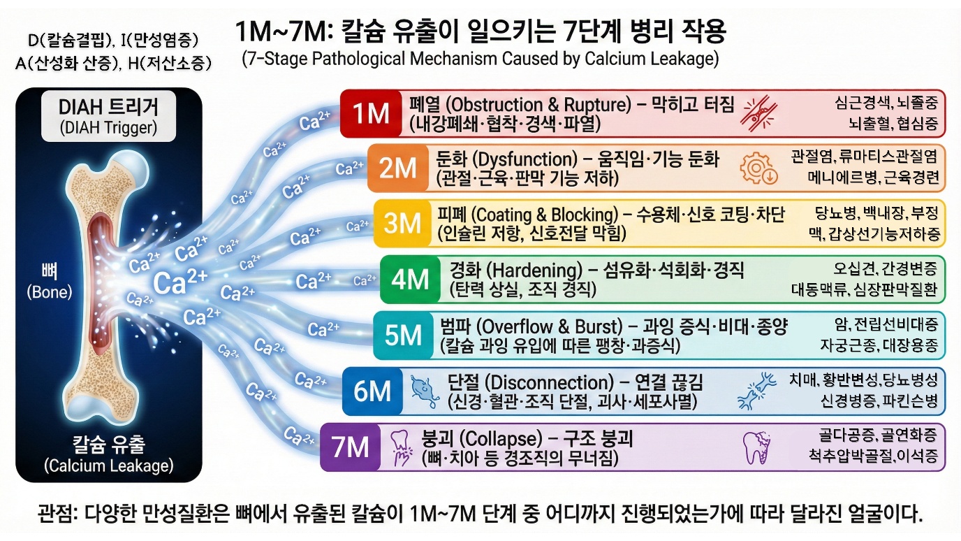
The first is 1M Obstruction & Rupture, blocking and bursting. When calcium deposits on the vessel wall, the lumen narrows, stenosis forms, blood flow is blocked and infarction occurs, and in severe cases the vessel ruptures and hemorrhage occurs. Myocardial infarction, stroke, and cerebral hemorrhage are examples of this pathway.
The second is 2M Dysfunction, growing dull. When calcium deposits on joints, muscles, and valves, movement grows dull and contraction and relaxation function decline. Degenerative arthritis, valve calcification, and heart failure are examples of this pathway.
The third is 3M Coating & Blocking, being coated and blocked. When calcium affects the receptors on the cell surface, signal transmission is blocked and resistance to hormones develops. The insulin resistance seen in type 2 diabetes may be related to this pathway.
The fourth is 4M Hardening, hardening. When calcium deposits in tissue, fibrosis and calcification proceed, and the tissue becomes rigid and loses elasticity. Arteriosclerosis, pulmonary fibrosis, and cirrhosis of the liver are examples of this pathway.
The fifth is 5M Overflow & Burst, overflowing and bursting. When calcium floods excessively into the cell, the cell hyperproliferates, becomes enlarged, and a tumor forms, and in the extreme case the cell expands, bursts, and dies. Cancer and benign prostatic hyperplasia may be related to this pathway.
The sixth is 6M Disconnection, being cut and severed. When calcium deposits on blood vessels or nerves, blood flow is completely blocked, nerve transmission is severed, and tissue undergoes necrosis. Diabetic foot, dementia, and Buerger's disease are examples of this pathway.
The seventh is 7M Collapse, falling apart. When calcium escapes excessively from the bones, the structure of hard tissue such as bone and teeth collapses. Osteoporotic fracture and tooth loss are examples of this pathway. From 1M to 6M are problems that arise from calcium piling up where it should not be, and 7M is a problem that arises from calcium escaping from where it should be. They are two sides of the same calcium imbalance. Chapter 7 deals with these mechanisms in detail.
The core of the DIAH-7M aging and chronic disease pathway framework is this. Chronic diseases appear individual, but they share a common pathway. At the center of that common pathway is calcium. The DIAH triggers cause calcium to flow out of the bones, and the calcium that has flowed out leads to chronic disease through the 7M mechanisms. That vitamin D deficiency is related to osteoporosis, that inflammation promotes bone loss, that acidosis weakens bone, and that vascular calcification is related to cardiovascular disease are all scientific facts established by countless studies. DIAH-7M is not a new discovery but an integrated framework that arranges puzzle pieces already revealed into a single picture. The reason this framework is useful is that it lets us see the chronic diseases that appear individual from a single viewpoint, and it presents an integrated approach to prevention and management. If osteoporosis and arteriosclerosis are not separate diseases but two sides of the same calcium imbalance, then the prevention strategy too must be integrated.
Conclusion
In this chapter we have learned the fact that, through the evolutionary background, DNA designs and calcium executes. From fertilization to apoptosis, calcium pulls the trigger at every decisive moment of life. Life cycles through six stages: birth, growth, reproduction, decline, death, and return. The key to each stage is calcium. In birth a calcium wave awakens life, in growth calcium fills the bones, in reproduction the calcium metabolism system is optimized, and in decline a double lock operates so that absorption is blocked and efflux is promoted. Decline may not be a mere breakdown but an exit that evolution designed. Antagonistic pleiotropy, the evolutionary trade-off in which a gene advantageous when young becomes harmful when old, is its mechanism. Too many systems change in the same direction. It is hard to see it as random.
In Chapter 6 we will look in detail at each mechanism of the DIAH triggers by which calcium operates as an execution code, and in Chapter 7 at each of the 7M pathological pathways.
References
1. Whitaker, M. (2006). Calcium at fertilization and in early development. Physiological Reviews, 86(1), 25-88. doi:10.1152/physrev.00023.2005
2. Bers, D. M. (2002). Cardiac excitation-contraction coupling. Nature, 415(6868), 198-205. doi:10.1038/415198a
3. Südhof, T. C. (2012). Calcium control of neurotransmitter release. Cold Spring Harbor Perspectives in Biology, 4(1), a011353. doi:10.1101/cshperspect.a011353
4. Khosla, S., Oursler, M. J., & Monroe, D. G. (2012). Estrogen and the skeleton. Trends in Endocrinology & Metabolism, 23(11), 576-581. doi:10.1016/j.tem.2012.03.008
5. Weitzmann, M. N., & Pacifici, R. (2006). Estrogen deficiency and bone loss: An inflammatory tale. Journal of Clinical Investigation, 116(5), 1186-1194. doi:10.1172/JCI28550
6. Williams, G. C. (1957). Pleiotropy, natural selection, and the evolution of senescence. Evolution, 11(4), 398-411. doi:10.2307/2406060
7. Kirkwood, T. B. L., & Austad, S. N. (2000). Why do we age? Nature, 408(6809), 233-238. doi:10.1038/35041682
8. Clapham, D. E. (2007). Calcium signaling. Cell, 131(6), 1047-1058. doi:10.1016/j.cell.2007.11.028
9. Marambaud, P., Dreses-Werringloer, U., & Vingtdeux, V. (2009). Calcium signaling in neurodegeneration. Molecular Neurodegeneration, 4, 20. doi:10.1186/1750-1326-4-20