
This article is the full text of Chapter 4 of The Declaration of the Age of Physical Medicine (Yoon Jong-won). It is an academic exposition presenting the author's hypothesis of physical medicine, and the body text, figures, and citations follow the original manuscript.
The roughly 1 kilogram of calcium contained in our body all came from the death of a star. Billions of years ago some star ended its life and exploded, and one of the heavy elements that explosion scattered into the universe was calcium. That calcium flowed through billions of years of time to form the coral and shells and animal bones of the Earth, to form the bones of our parents, and at this very moment the remnant of that star is flowing within the body of the reader reading this text. We are all the remnants of a star.
And yet this element that came from a star is the most decisive executing code of every living organism. Let us recall that first moment when a person's life begins. Modern medicine has already revealed what the first molecular signal is by which, at the moment the sperm and the egg meet, those two cells combine and a new life begins. It is the explosive influx of calcium. The calcium concentration inside the egg momentarily surges up, and that first calcium signal ignites all the subsequent events of development. If that signal does not turn on, no life begins at all. A person's life begins with the ignition of a calcium signal.
Here we must note one decisive fact. In the latter half of the 19th century, Darwin, through natural selection, gave us the answer to who evolves. In the mid-20th century, Watson and Crick, through the discovery of the double helix, gave us the answer to what evolves. But one decisive question remained empty. Who on earth executes that who and that what? Which molecule turns on the first signal, and which molecule turns off the last signal? How does the information written in DNA become a living event? What fills that empty seat is precisely calcium. Calcium is the executor of life.
Let us recall one analogy. For a symphony to resound, three things are needed. The score, the conductor, and the players. With the score alone, the music does not begin. Even if all the members are holding their instruments and sitting in their seats, if the conductor does not give the first gesture, not a single note resounds. Music is not heard by looking into the score; it begins to be heard only when the conductor executes that score. The molecular events of life have exactly the same structure. DNA is the score that writes down what must be done. The cell is the player holding that score. And calcium is the conductor that turns that score into actual music. That calcium which floods in explosively at the moment the sperm and the egg meet is the conductor's first gesture announcing the beginning of the symphony that is one human being. Without that gesture, no score, and no player, can make a single note of music.
[Figure 1] Music and Life, Two Landscapes of the Same Structure
| Element of music | Role in music | Element of life | Role in life |
|---|---|---|---|
| Score | What must be done | DNA | Which protein must be made |
| Player | Actually performs that "what" | Cell | Actually performs that "what" |
| Conductor | Begins with the first gesture, controls the whole flow | Calcium | Begins with the first signal, mediates the whole flow |
Even after that first gesture, calcium executes every decisive moment of a person's life. When the fetus's bones are made, calcium is deposited at that place. When a child grows and gains height, calcium accumulates at the ends of the bones. When the hormone secretion of puberty begins, calcium mediates that signal. When a young person's muscle contracts, when a nerve transmits a signal, when an immune cell recognizes an intruder, calcium is present at every moment. And even at the moment of reproduction, calcium mediates. That first signal that began in the union of the sperm and the egg is ignited again at the beginning of the next generation. Right up to the moment a person makes the first life of the next generation, calcium executes the code without fail at every stage.
And yet, viewed on the time scale of evolution, one decisive fact is revealed. It is that evolution has never designed the eternal survival of the individual. What evolution designed is only one thing, the transmission of genes through reproduction. When an individual reaches the stage of reproduction and leaves the next generation, that individual's evolutionary mission is essentially over. From evolution's standpoint, the time thereafter is merely an auxiliary period until the superior genes are safely established in the next generation. So all the intricate homeostatic systems of the human body are designed with the reproductive-age years as the peak, and the human body after reproduction enters a period in which that system slowly moves to another stage.
And precisely at this post-reproductive stage, calcium begins to show a new face. To be precise, the same calcium begins to execute a different stage of the same code. The calcium that operated as the ignition signal of life in the stages of birth and growth and reproduction operates as the medium of deposition in the stages of decline and death. It is the same conductor conducting a different movement of the same symphony. If the first movement was the fast tempo of ignition, the last movement is the slow tempo of sinking. The essence of the change that our parents' generation begins to face after their fifties lies here.
[Figure 2] The Role of Calcium by Life Stage
| Stage | Period | Role of calcium | Substance |
|---|---|---|---|
| Birth | Sperm-egg union | Ignition of the developmental signal | First calcium burst |
| Growth | Childhood to youth | Mediation of bone, muscle, nerve, immunity | Normal medium |
| Reproduction | Youth to middle age | Mediation of the next generation's first signal | Next-generation ignition |
| Start of decline | After the fifties | Mediation of the crisis emergency prescription | Withdrawal from bone begins |
| Progressive deposition | Sixties to seventies | Lingers in microvasculature, arteries, soft tissue | Cannot return to its own place |
| Death | The last stage | The last mediation of the emergency prescription | Passage through the four gateways |
In fact, even wrinkles, the most everyday sign of aging, are not unrelated to the code execution of calcium. As the elastic fibers of the skin weaken and collagen degenerates, minute traces of calcium begin to settle there together, and the skin gradually loses its original elasticity. The event of calcium accumulating in the artery and hardening it and the event of calcium lingering together at the place where the skin's elasticity has collapsed are the execution of one and the same code, differing only in location. The wrinkles deepening on our parents' faces, the chronic diseases accumulating in their bodies, the aching joints, and the hardening arteries are all the surfacing, at different places, of one and the same calcium code.
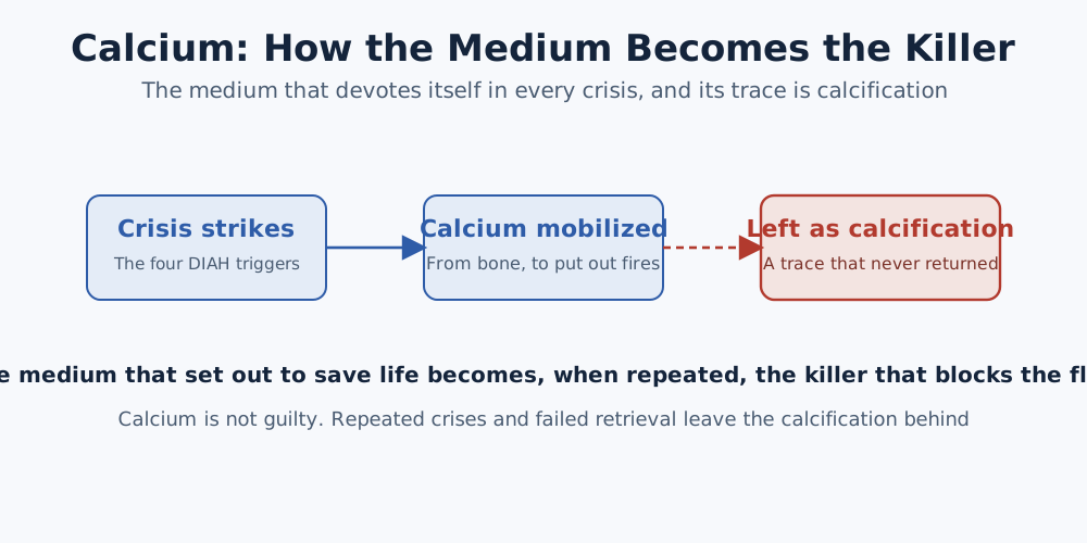
Here there is something to make clear. Calcium is by no means a bad substance. One must not view calcium as an enemy or fear it. Rather, calcium is the victim. Whenever the human body meets a crisis, calcium leaves its own place voluntarily. When deficiency strikes, it comes out of the bone to protect the blood concentration. When inflammation occurs, it is mobilized to awaken the immune cells. When acidosis begins, it comes out together to neutralize the acid. When hypoxia strikes, it enters to turn on the adaptation signal. In every crisis it steps forward first and gives itself first. The human body calls calcium in order to live, and calcium answers that call. But when the crisis becomes an everyday thing and the call continues endlessly, calcium cannot return to its own place. Called out too often, it has no time to return to the bone again. So it lingers on the microvascular wall, in the artery, in the soft tissue, in the skin. There the deposition begins. What we call calcification is in fact the trace of calcium that could not return to its own place. Our parents' hardened arteries, aching joints, and deepening wrinkles are not events created out of hatred for calcium. They are the trace of calcium that came out of its own place to quell the small crises our parents cried out for day after day throughout the years they lived, and that could not all return. The calcification in our parents' bodies is not something hateful, but evidence of the years our parents lived protecting their family, and the mark of calcium's devotion to protect those parents. Calcium is not the enemy. Calcium is the victim. And the trace of that sacrifice is calcification.
That the same molecule has different faces according to place and flow, this is the ambivalence of calcium. And this ambivalence is, before any molecular-dimension chemical reaction, a physical event of flow. When it flows it is a medium, and when it stops it is a sediment. This is the reason this book bears the name physical medicine. This chapter is the story of that ambivalence. It unravels in turn how the same calcium operates simultaneously as the medium of life and the medium of deposition within the body of the same person, and what condition determines those two faces. It is not to fear calcium. It is to clearly understand what calcium did for us, and what we did to calcium.
Calcium: Its Original Role as the Medium of Life
The body of an adult contains about 1 kilogram of calcium. Of that, about 99 percent is contained in the bones and teeth, and the remaining 1 percent is distributed in the blood, the extracellular fluid, and inside cells. It is a small proportion of 1 percent, but this 1 percent mediates almost every decisive event of the activity of life.
[Figure 3] Distribution of Calcium in the Human Body
| Location | Proportion | Main form | Role |
|---|---|---|---|
| Bone and teeth | About 99% | Hydroxyapatite crystal | Skeletal support, calcium storehouse |
| Blood, extracellular fluid | About 1% | Ionic calcium, bound calcium | Medium transport pathway |
| Intracellular | About 0.01% | Free calcium, stored calcium | The main body of signal transduction |
As the distribution above shows, 99 percent is stored in the bone and the rest is distributed in the blood and cells. And the amount of calcium inside the cell is very small, at about the 0.01 percent level. But this trace intracellular calcium operates as the most decisive signal of every cell.
The activity of life that calcium mediates is wide-ranging. It can be organized into five core roles.
[Figure 4] The Five Medium Roles of Calcium
| Role | Site of operation | Representative event |
|---|---|---|
| Muscle contraction | Cardiac, skeletal, smooth muscle | Heartbeat, exercise, blood pressure regulation |
| Neurotransmission | Neuron synapse | Thought, sensation, motor commands |
| Secretion | Secretory cells | Insulin, hormones, digestive enzymes |
| Blood coagulation | Site of vessel damage | Hemostasis |
| Division and death | All cells | Cell regeneration, apoptosis |
First, muscle contraction. That the heart beats once per second is also mediated by calcium. When the calcium concentration momentarily rises inside a myocardial cell the muscle contracts, and when the concentration drops again the muscle relaxes. The movement of skeletal muscle, the peristalsis of gastrointestinal smooth muscle, and the contraction and relaxation of vascular smooth muscle are all mediated by calcium. Without a calcium signal, not even a single heartbeat occurs.
Second, neurotransmission. When a signal arrives at the end of a neuron the calcium channel opens, and as calcium enters it triggers the secretion of neurotransmitters. Calcium is present among the molecular-level signals of every process by which we think, feel, and make decisions.
Third, secretion. That insulin is secreted, that hormones are secreted, and that digestive enzymes are secreted all begin with a calcium signal. Inside a secretory cell, a minute change in calcium concentration serves as the last signal that fuses the secretory vesicle with the cell membrane.
Fourth, blood coagulation. The coagulation cascade, in which coagulation factors are activated in turn when a vessel is damaged, requires calcium at several stages. Without calcium the blood does not clot. This is precisely why a calcium chelator is put into a drawn-blood tube to prevent coagulation.
Fifth, cell division and death. When a cell divides, and when a cell decides on death, the calcium signal plays a decisive role. Calcium is present at every decisive moment of a cell's lifetime, that is, from birth to death.
The reason calcium can mediate all these roles is that calcium is regulated very precisely at a very low concentration. The serum calcium concentration is maintained within the very narrow range of 8.8 to 10.4 mg/dL. The calcium concentration inside the cell is more than about ten-thousand times lower than that. This ten-thousand-fold concentration difference is the decisive condition for it as a medium. It is a structure in which even a small change of concentration can create a large change of signal.
The Ambivalence of Calcium: Flow and Sediment
For calcium to operate as a medium, two conditions are needed. First, a very low concentration. Second, ceaseless flow.
Concentration is measured well by medicine through tests. If the serum calcium in a blood test is within the normal range, we are reassured. But even if the concentration is normal, if the flow stops, calcium can change from a medium into an enemy.
To understand this, let us recall one analogy. When a river flows, sand is mixed into the river water and carried. But when the river loses its flow and begins to stagnate, the sand begins to pile up on the bottom. The same sand plays a different role according to the presence or absence of flow. When it flows, a carried medium; when it stops, a piled-up sediment.
Calcium is likewise. Flowing calcium transmits signals, contracts muscles, and triggers secretion. Stopped calcium begins to precipitate. The same molecule plays two roles according to location and flow. We call this property the ambivalence of calcium. A molecule that can be both a medium and an enemy.
This ambivalence originates in the chemical characteristics of calcium. Calcium is a cation with two positive charges. Because the positive charge is strong, it has a tendency to bind strongly with negatively charged molecules (phosphate groups, proteins, lipids). When the flow is sufficient, this binding is temporary and reversible. It mediates signals while repeating binding and releasing. But when the flow weakens and calcium lingers long in one place, the binding stabilizes and a crystal is formed. The most stable crystal that calcium and phosphate make by binding is the mineral called hydroxyapatite. The same mineral is also the main component of bone and teeth.
[Figure 5] Medium Calcium and Enemy Calcium: A Comparison of Ambivalence
| Comparison item | Medium calcium | Enemy calcium |
|---|---|---|
| State | Flowing ion (Ca²⁺) | Settled crystal (hydroxyapatite) |
| Location | Blood, extracellular fluid, intracellular | Microvascular wall, space between tissue |
| Binding | Temporary, reversible | Stable, irreversible |
| Action | Signal transduction, muscle contraction, secretion | Flow cutoff, inflammation induction |
| Retrieval | Returns to its own place | Cannot return |
In the bone this crystal is normal. Bone is a tissue in which calcium and phosphate are deposited as crystals to create hardness. But when the same crystal is made in a place other than bone, that is abnormal. When a crystal is made inside the microvascular wall, the flow is blocked; when a crystal is made between the joints, pain arises; and when a crystal is made in the kidney tubule, it becomes a kidney stone. The crystal itself is the same. Only the location differs.
Here a decisive insight emerges. That calcium changes from a medium into an enemy is not that calcium changed but that the flow changed. Calcium retains its own original nature just as it is. Only, at the place where the flow has weakened, that original nature creates a different result.
This insight raises the view of aging and chronic disease by one stage. Reducing the calcium itself that comes in from natural diet is not the answer. Making the flow alive is the answer. When the flow is alive, the same calcium operates as a medium. When the flow weakens, the calcium that has come out of the bone begins to settle in another place. The calcium that becomes an enemy is not newly introduced calcium from outside, but calcium that has taken the wrong place within the human body. This is not a problem that is solved by reducing calcium. It is a problem that is solved by reviving the flow.
From Medium to Enemy: The Mechanism of Conversion
There is an exact sequence in the process by which calcium is converted from a medium to an enemy. Following that sequence yields the molecular-level picture of how aging and chronic disease begin.
The first stage is the emergency withdrawal. When the human body senses a signal of calcium deficiency, or acidification, or inflammation, parathyroid hormone is secreted. Parathyroid hormone sends a signal to the bone and activates the bone's osteoclasts. The osteoclasts dissolve the mineral of the bone and release calcium and phosphate into the blood. In the emergency period, the calcium storehouse of the bone is released. Because the human body must maintain the blood calcium concentration within a narrow range, it operates this emergency withdrawal at any cost.
What regulates this emergency withdrawal is the balance of three hormones.
[Figure 6] The Triple Axis of Calcium-Regulating Hormones
| Hormone | Secretion condition | Action | Result |
|---|---|---|---|
| Parathyroid hormone | Blood calcium ↓ | Withdrawal of calcium from bone, kidney reabsorption ↑ | Blood calcium ↑ |
| Calcitonin | Blood calcium ↑ | Osteoclast suppression, decreased bone resorption | Blood calcium ↓ |
| Vitamin D (active form) | Sunlight, diet | Calcium absorption in the intestine ↑ | Blood calcium ↑ |
As the figure shows, the three hormones, parathyroid hormone, calcitonin, and vitamin D, operate as one system. When parathyroid hormone withdraws calcium from the bone, calcitonin induces that calcium to be stored in the bone again, and vitamin D absorbs new calcium in the intestine and replenishes the balance of the whole system. This intricate triple axis maintains the ordinary blood calcium concentration within a very narrow range. But in a chronic crisis situation, this triple axis tilts to one side. The withdrawal accelerates, the storage cannot keep up, and absorption alone cannot fill the withdrawal amount. The calcium released that way passes to the next stage.
The second stage is the fate of the released calcium. In a normal situation, the released calcium returns to the bone again after finishing its medium role. Calcitonin and vitamin D regulate this retrieval process. But in an environment where chronic deficiency, inflammation, acidification, and hypoxia persist, parathyroid hormone is chronically activated and the emergency withdrawal does not stop. The speed at which the released calcium is retrieved back into the bone cannot keep up with the speed at which it is withdrawn. Calcium begins to accumulate in the blood and the space between tissue.
The third stage is the appearance of a place to settle. When the microvascular wall is damaged, or inflammation accumulates in the space between tissue, or the extracellular matrix is deformed, a negatively charged surface on which calcium can settle is made. The membrane of a damaged endothelial cell, the vesicles left by a dead cell, degenerated collagen. All these surfaces become nucleation sites where calcium and phosphate can begin to crystallize.
The fourth stage is crystal formation. When calcium and phosphate gather at the nucleation site, at first an amorphous calcium-phosphate precipitate is made. As time passes this precipitate grows into a crystal structure and finally becomes a hydroxyapatite crystal. It is the same crystal as bone, but this time a crystal made in a place other than bone.
The fifth stage is active calcification. A key study published by the Reynolds group of Cambridge University in the UK in the Journal of the American Society of Nephrology made one decisive fact clear. It is that when the calcium and phosphate concentration rises, vascular smooth muscle cells begin to secrete small membrane vesicles, and inside those vesicles calcium and phosphate grow into crystals. It is that the vascular cell is not merely passively exposed to calcium, but is converted into a cell that actively makes the place of crystal formation. It is that the vessel of one organ behaves as if it were beginning to make a small bone. A comprehensive review published by the Demer-Tintut group of the University of California in the U.S. cardiovascular journal Circulation organized the essence of this event. It is that vascular calcification is not a simple aging byproduct but an intricate pathological process that uses the molecular circuit of bone formation. The same molecular signal, the same crystal-formation pathway, operates as normal in the bone and as pathology in the vessel.
It would be good if one could stop it anywhere in the five stages. But as long as deficiency, inflammation, acidification, and hypoxia persist chronically, the five stages progress slowly but ceaselessly. Active calcification, once begun, has a self-reinforcing loop, and it accelerates with time.
Microcalcification: The Identity of the New Enemy
The product that the calcium converted from medium to enemy creates is precisely microcalcification. Microcalcification is a micro-dimensional precipitate different from general calcification.
[Figure 7] Comparison of Microcalcification and Large-Vessel Calcification
| Item | Microcalcification | Large-vessel calcification |
|---|---|---|
| Size | Nanometer to micrometer | Millimeter to centimeter |
| Location | Microvascular wall, space between tissue | Large artery wall |
| Imaging test | Not detected by ordinary CT/MRI | Detectable by CT/MRI |
| Distribution | Systemic, multiple | Localized, focal |
| Reversibility | Does not self-dissolve | Some drug effect |
In terms of size, microcalcification is at the nanometer to micrometer unit. Because the spatial resolution of ordinary medical imaging is about 0.5 millimeters or more for computed tomography, microcalcification is not caught by imaging. While everything comes back normal on the patient's test results, at the micro dimension the crystal deposition progresses.
In terms of location, microcalcification is distributed on the inside of the microvascular wall and in the space between tissue. The calcification of a large artery is caught by imaging as atherosclerosis, but the calcification of the microvasculature occurs at a dimension smaller than that, so it is not caught. But the total length of the microvascular network is overwhelmingly longer than the large vessels, and because all cells depend on the microvasculature, the clinical impact that micro-dimensional deposition creates is overwhelmingly large relative to its size.
In terms of chemical composition, microcalcification has as its main component hydroxyapatite, in which calcium and phosphate are bound. It is the same mineral as bone. But unlike bone, microcalcification cannot dissolve or be absorbed on its own. Microcalcification, once formed, remains at that place and accumulates. With time the amount of deposition increases, and the bidirectional exchange of the microvasculature progressively weakens.
In terms of action, microcalcification does not stop at merely narrowing the pathway. Microcalcification itself creates a new inflammation signal. Immune cells recognize microcalcification as an external intruder and start an inflammatory response. The inflammation that starts induces more calcium withdrawal, more calcium makes more microcalcification, and more microcalcification makes stronger inflammation. The self-reinforcing loop accelerates progressively.
This is the true underlying reality of aging and chronic disease. Before the blockage of the large vessels, the microcalcification deposition progressing slowly but surely inside the microvasculature. That event specialty medicine did not see. That deposition progressing even while imaging comes back normal. It is one and the same event occurring simultaneously in multiple organs within the body of the same person.
[Figure 8] A Self-Check of Calcium Ambivalence
| Your calcium: is it a medium, or an enemy? Please check whether two or more of the following items applied to you over the past year. 1. Even though you usually take in enough calcium through natural diet such as anchovies, your bone density test result is not good. 2. Your serum calcium concentration comes back normal in a blood test, but you were diagnosed with osteoporosis. 3. You have been told of a finding of the beginning of atherosclerosis in a vascular test. 4. Since your fifties, your hands and feet have grown cold more often. 5. There is someone in your family who was diagnosed with osteoporosis and cardiovascular disease together. If two or more items apply to you, there is a strong possibility that the ambivalence of calcium has already begun to operate. It is a signal that the polarization in which calcium leaves the bone while at the same time calcium is deposited in the vessel, that is, the calcium paradox, has begun. The following chapters address what starts and accelerates this polarization. |
|---|
We have examined how calcium changes from a medium into an enemy. But what starts this conversion? What signal makes the human body begin the emergency withdrawal?
The answer is a combination of four signals. Deficiency, inflammation, acidification, hypoxia. When these four signals combine and issue the emergency command to the human body, calcium begins to convert from a medium into an enemy. Binding the English initials of these four signals, we call it DIAH. The next chapter is the story of those four triggers.
References
[1] Berridge, M. J. (2012). Calcium signalling remodelling and disease. Biochemical Society Transactions, 40(2), 297-309.
[2] Brown, E. M., & MacLeod, R. J. (2001). Extracellular calcium sensing and extracellular calcium signaling. Physiological Reviews, 81(1), 239-297.
[3] Clapham, D. E. (2007). Calcium signaling. Cell, 131(6), 1047-1058.
[4] Demer, L. L., & Tintut, Y. (2008). Vascular calcification: Pathobiology of a multifaceted disease. Circulation, 117(22), 2938-2948.
[5] Hofbauer, L. C., Brueck, C. C., Shanahan, C. M., Schoppet, M., & Dobnig, H. (2007). Vascular calcification and osteoporosis: from clinical observation towards molecular understanding. Osteoporosis International, 18(3), 251-259.
[6] Orrenius, S., Zhivotovsky, B., & Nicotera, P. (2003). Regulation of cell death: The calcium-apoptosis link. Nature Reviews Molecular Cell Biology, 4(7), 552-565.
[7] Peacock, M. (2010). Calcium metabolism in health and disease. Clinical Journal of the American Society of Nephrology, 5(Suppl 1), S23-S30.
[8] Reynolds, J. L., Joannides, A. J., Skepper, J. N., McNair, R., Schurgers, L. J., Proudfoot, D., Jahnen-Dechent, W., Weissberg, P. L., & Shanahan, C. M. (2004). Human vascular smooth muscle cells undergo vesicle-mediated calcification in response to changes in extracellular calcium and phosphate concentrations: A potential mechanism for accelerated vascular calcification in ESRD. Journal of the American Society of Nephrology, 15(11), 2857-2867.
[9] Thompson, B., & Towler, D. A. (2012). Arterial calcification and bone physiology: Role of the bone-vascular axis. Nature Reviews Endocrinology, 8(9), 529-543.