
This piece is the first part of Chapter 4 of Calcium: The Reality of Evolution That Darwin and the Genome Map Missed (Yoon Jong-won). The body, figures, and citations follow the manuscript as written.
Introduction: How 86 Billion Neurons Talk to One Another
The human brain has about 86 billion neurons. This number, measured precisely in 2009 by the Brazilian neuroscientist Suzana Herculano-Houzel using a new methodology, revised the estimate of 100 billion that had been printed in textbooks until then.
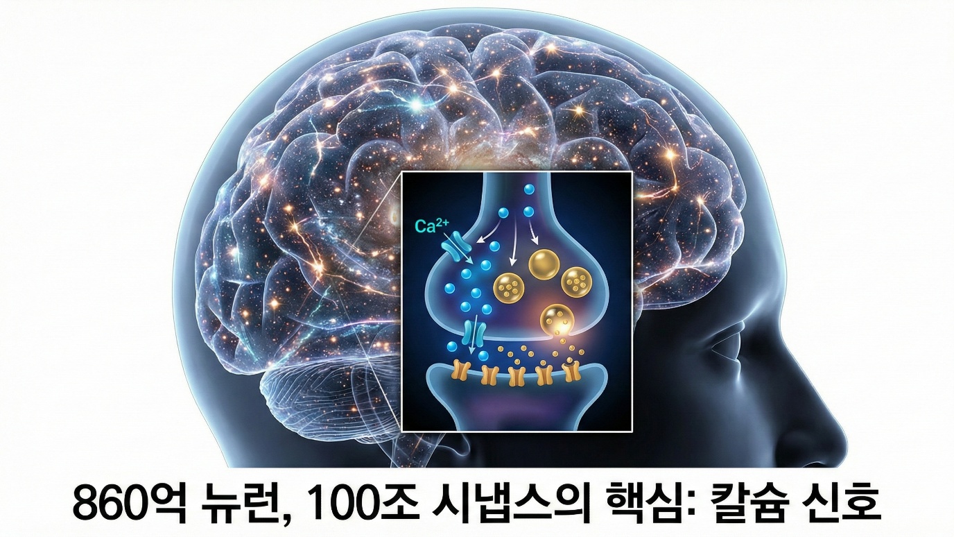
The difference between 86 billion and 100 billion may sound trivial, but that difference of 14 billion is a number equal to the total neuron count of an entire baboon brain, so it is by no means a small error. And these 86 billion neurons are connected to one another by about 100 trillion synapses. It is hard to grasp the number 100 trillion, but since the number of stars in our galaxy is estimated at about 100 billion to 400 billion, the number of synapses in the human brain is several hundred times greater than the stars of our galaxy. On average each neuron forms about 1,000 or more synapses, and at every moment hundreds of billions of signals pass back and forth across this vast network. Every time we see, hear, feel, think, and remember something, these signals flow, and in your own brain reading this very sentence, hundreds of billions of signal transmissions are taking place at this very moment. And at the heart of all these signals is that ion this book has tracked from the very beginning: calcium.
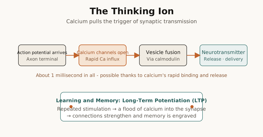
Signal transmission between one neuron and another, that is, synaptic transmission, is impossible without calcium. When an action potential is transmitted along the axon and reaches the synaptic terminal, voltage-gated calcium channels open and calcium ions push their way into the cell, and this calcium fuses the vesicles holding neurotransmitters with the cell membrane and makes them release the neurotransmitters. This process, which takes place within several hundred microseconds to a few milliseconds, makes all communication among the 86 billion neurons possible, and every function of the brain is built upon this basic process. Learning and memory likewise depend on calcium. The core signal of the phenomenon in which a synapse is strengthened or weakened, that is, synaptic plasticity, is calcium itself: when the calcium concentration is high the synapse is strengthened and long-term potentiation occurs, and when the calcium concentration is low the synapse is weakened and long-term depression occurs. But here the familiar paradox that runs through this book appears again. In Chapter 1 we learned that calcium is toxic to the cell. That it is a dangerous ion that precipitates ATP, aggregates proteins, destroys the cell membrane, and in the end kills the cell. In the brain in particular, calcium overload brings about the death of nerve cells, and this is excitotoxicity, the main cause of the large-scale neuronal damage that follows a stroke. Signal and poison. Calcium performs these two roles at once in the brain. How did the evolution of higher intelligence resolve this paradox? In this chapter we will look at what role calcium plays in brain function, and at how the refinement of the ability to control calcium became the material foundation of higher intelligence.
The Synapse: Calcium Pulls the Trigger
In 1967, Bernard Katz and Ricardo Miledi of University College London made a groundbreaking discovery at the neuromuscular junction. They were studying signal transmission between nerve and muscle using a frog neuromuscular preparation, and they carried out a simple but decisive experiment. When they removed calcium from the solution in which the preparation was immersed, the muscle did not contract even when the nerve was stimulated. The nerve transmitted the action potential normally, and the muscle too was capable of contracting when stimulated directly, but the signal was not transmitted from nerve to muscle. Yet when calcium was put back into the solution, nerve transmission returned to normal. Even more decisive was the timing. Signal transmission occurred only when calcium was present at the same time as the nerve stimulation or just before it, and adding calcium after the stimulation was of no use at all. This meant that calcium was the direct trigger of neurotransmitter release. Without calcium, no matter how strongly the nerve was activated, no neurotransmitter was released, and therefore no signal was transmitted to the next cell. For this discovery Katz received the Nobel Prize in Physiology or Medicine in 1970, and more than half a century of research since has confirmed again and again, at the molecular level, that calcium is the key trigger of synaptic transmission.
Today we know in astonishingly fine detail how synaptic transmission takes place. The action potential generated in the cell body of the neuron is transmitted along a long projection called the axon and reaches the synaptic terminal. Embedded in the cell membrane of the synaptic terminal is a special protein called the voltage-gated calcium channel, which is normally closed but opens when the membrane potential is depolarized, that is, when the action potential arrives. The instant the channel opens, calcium ions push their way from outside the cell to inside it. As we saw in Chapter 1, the calcium concentration outside the cell is about 1 to 2 millimolar and the calcium concentration inside the cell is about 100 nanomolar, so the concentration difference is more than ten thousand-fold. Because of this enormous concentration gradient, when the channel opens calcium flows in rapidly, as water pours out when the floodgate of a high dam is opened. The calcium that flows in binds to a calcium-sensing protein called synaptotagmin, and synaptotagmin sits attached to the surface of the vesicles holding neurotransmitters, waiting for calcium to come. When calcium binds to synaptotagmin the structure of this protein changes, and this structural change activates a protein machine called the SNARE complex. The SNARE complex forcibly pulls the vesicle membrane and the cell membrane close together and fuses them, and when fusion occurs the neurotransmitters held inside the vesicle are released into the synaptic cleft, the narrow space between the two neurons. The released neurotransmitters reach the receptors of the postsynaptic neuron through diffusion and bind to them, and this gives rise to a new electrical signal in the postsynaptic neuron.
The most astonishing thing in this whole process is the speed. It takes only several hundred microseconds from the opening of the calcium channel to the beginning of vesicle fusion, and the whole process from the release of neurotransmitters to the appearance of an electrical signal in the postsynaptic neuron is completed within a few milliseconds. A millisecond is one thousandth of a second and a microsecond is one millionth of a second. Since it takes us about 300 milliseconds to blink, several hundred synaptic transmissions can occur during a single blink of the eye. How is such a fast process possible? The answer lies in spatial proximity. In a special zone of the synaptic terminal called the active zone, the calcium channels and the neurotransmitter vesicles are arranged very close together. The distance between them is only a few tens of nanometers, which is a distance corresponding to a few thousandths of the thickness of a hair. When calcium comes out of the calcium channel it can immediately reach the synaptotagmin of the vesicle right beside it. In this narrow space the calcium concentration shoots up in an instant to several tens of micromolar, which is several hundred times the basal concentration of 100 nanomolar. The principle we saw in Chapter 1 works here too. Because the basal concentration is extremely low, the relative change when a signal comes is dramatic, and the signal is clear. And because calcium pumps and calcium-binding proteins remove calcium rapidly, the signal terminates abruptly and is ready to receive the next signal. Low basal concentration, sharp rise, rapid termination. Calcium is excluded because it is a poison, and because it is excluded it becomes a clear signal. This paradox works in the same way at the synapse as well, and it is repeated hundreds of billions of times at every moment across the 100 trillion synapses among 86 billion neurons.
Learning and Memory: Calcium Changes the Synapse
How does the brain learn and remember? This question is a puzzle that has fascinated philosophers and scientists for thousands of years. The most compelling answer in neuroscience today is synaptic plasticity. Synapses are not fixed; depending on the pattern of use they grow stronger and grow weaker. The synapses between neurons that are frequently activated together are strengthened, and the synapses between neurons that are not activated together are weakened. This is the principle proposed by the Canadian psychologist Donald Hebb in his book "The Organization of Behavior," published in 1949. Hebb offered a theoretical conjecture about what happens in the brain when learning occurs, and the heart of it was this. When two neurons are repeatedly activated together, the connection between them is strengthened. Summarized in English by the adage "Neurons that fire together, wire together," that is, neurons that fire together become wired together, this principle became one of the most famous ideas in neuroscience. But when Hebb proposed this idea it was a purely theoretical conjecture, and there was no experimental evidence that synapses are actually strengthened.
In 1973, Terje Lømo of Norway and Tim Bliss of Britain found experimental evidence for this principle in the hippocampus of a rabbit. The hippocampus is a small structure located deep in the temporal lobe of the brain that plays a central role in the formation of new memories. A patient whose hippocampus is damaged cannot form new memories. Lømo and Bliss repeatedly gave high-frequency electrical stimulation to a particular neural pathway in the rabbit hippocampus. It was typically a rapid stimulation of about 100 times per second given for one second. Remarkably, after this high-frequency stimulation was given, the synaptic response of that pathway was strengthened, and this strengthening lasted for several hours, even several days. A stronger response came out even when a stimulus of the same intensity was given. It seemed as if the pathway had grown stronger through being used. This phenomenon is called long-term potentiation, Long-Term Potentiation in English, LTP for short. Long-term potentiation was the experimental reality of the synaptic strengthening that Hebb had predicted, and it came to be regarded as the cellular basis of learning and memory. And decades of research since then have revealed that the core signal of long-term potentiation is calcium itself.
What plays an important role in inducing long-term potentiation is a special protein called the NMDA receptor. The NMDA receptor is a receptor for glutamate, an excitatory neurotransmitter, and it has a very peculiar property, so much so that it is called a device that implements Hebb's principle at the molecular level. An ordinary ion channel, the AMPA receptor for example, opens immediately when a neurotransmitter binds to it. When glutamate comes the channel opens and ions flow. But the NMDA receptor is different. Even when glutamate binds, the channel does not open right away. This is because a magnesium ion is blocking the mouth of the channel. To release this magnesium block, the cell membrane must be depolarized. When the membrane potential rises sufficiently, magnesium leaves the channel, and only then does the NMDA receptor to which glutamate has bound open. In other words, for the NMDA receptor to open, two conditions must be met at the same time. The presynaptic neuron must be activated and release glutamate, and at the same time the postsynaptic neuron must also be activated by other inputs and be in a depolarized state. Only when the two neurons are activated together does the NMDA receptor open. Hebb's principle that neurons must fire together to be wired together is implemented at the molecular level. When the NMDA receptor opens, calcium ions flow into the postsynaptic neuron, and this calcium activates a series of signal transduction pathways that strengthen the synapse.
What is interesting is that the direction of synaptic change turns out to be exactly opposite depending on the calcium concentration. A high calcium concentration induces long-term potentiation. When the calcium concentration is high, calcium/calmodulin-dependent protein kinase II, called CaMKII, is activated. This enzyme is activated when calcium and calmodulin bind, and once activated it phosphorylates itself and maintains its active state for a while even after the calcium has disappeared. Activated CaMKII inserts more AMPA receptors into the postsynaptic membrane. Since the AMPA receptor is an ion channel opened by glutamate, when it becomes more numerous the synapse responds more strongly to the same amount of glutamate. The synapse is strengthened. Conversely, a low calcium concentration induces long-term depression, LTD for short. When the calcium concentration is low, instead of CaMKII another enzyme called calcineurin is activated. Calcineurin is a phosphatase, and it does the opposite of what CaMKII does. When calcineurin is activated, AMPA receptors are removed from the synapse and the synapse is weakened. The same signal, calcium, produces exactly opposite effects depending on its concentration. This means that the synapse is not simply strengthened alone but can be regulated in both directions according to the pattern of activity. Learning includes not only strengthening important connections but also weakening unnecessary ones, and calcium is the core signal of this bidirectional regulation.
The calcium signal induces not only short-term change but also long-term change. For synaptic strengthening to last beyond several hours or days to several months, several years, even a lifetime, something more permanent must change. A high calcium concentration activates a transcription factor called CREB. After being activated in the cytoplasm, CREB enters the nucleus and binds to DNA to promote the expression of particular genes. These genes synthesize new proteins, and the synthesized proteins are carried to the synapse and bring about structural changes in the synapse. The postsynaptic spines grow larger, and new synapses are formed. The very shape of the neuron changes. That this process is essential to the formation of long-term memory has been confirmed by many experiments. When a drug that blocks protein synthesis is administered, short-term memory forms normally but long-term memory does not form. Only when new proteins are synthesized during the several hours right after learning is that memory permanently stored. From calcium to CREB, from CREB to gene expression, from gene expression to protein synthesis, from protein synthesis to structural change. This pathway converts a fleeting experience into a permanent memory, and everything begins from the calcium signal. Of course there is still debate over whether synaptic plasticity is the sole storage mechanism of long-term memory, and some studies raise the possibility that other auxiliary mechanisms at the cellular level or the network level work alongside it. But modern neuroscience still sees little disagreement in regarding calcium-dependent synaptic plasticity as the central axis of memory formation.
The Brain's Paradox: Between Signal and Poison
If calcium is essential to neural signaling and learning, would it not be better to have more of it? Intuitively one might think so, but the reality is exactly the opposite. Calcium overload kills nerve cells. This is the phenomenon in which the toxicity of calcium we saw in Chapter 1 appears most dramatically in the brain, and it is called excitotoxicity. When a stroke occurs, a blood vessel is blocked or bursts and the blood supply to part of the brain is cut off. Cells starved of oxygen and glucose cannot make ATP in their mitochondria. In Chapter 1 we learned that the cell uses a considerable portion of its total energy to run the calcium pumps. Without ATP the calcium pumps stop working, and the cell can no longer pump calcium out. At the same time, glutamate is released excessively from the damaged cells suffering from the energy shortage. Under normal conditions glutamate release is finely regulated, but when energy is exhausted this regulation collapses. Because the pump that reabsorbs glutamate also requires ATP, the released glutamate accumulates in the synaptic cleft.
Excessive glutamate excessively activates the NMDA receptors of adjacent neurons. Under normal conditions the activation of NMDA receptors causes the calcium influx essential to learning, but excessive activation brings about catastrophe. Calcium flows in in massive amounts, and these cells too are already short of ATP and cannot pump out the calcium that has flowed in. Calcium overload begins. The massively influxed calcium produces all the toxic effects described in Chapter 1. The mitochondria try to absorb and buffer the calcium, but in an overloaded state the mitochondria themselves are damaged. Holes open in the mitochondrial membrane, a protein called cytochrome c is released and triggers the caspase cascade, and the cell death program is activated. At the same time, reactive oxygen species are generated in massive amounts from the damaged mitochondria and attack the molecules inside the cell. Proteins are oxidized, lipid membranes are damaged, and DNA is destroyed. The result is large-scale neuronal death. From the dying cells glutamate is released again and attacks the surrounding cells, and the chain reaction spreads. A considerable part of the neural damage after a stroke is caused not by the initial blockage of blood flow itself but by the excitotoxicity that follows. This is why time is so important in the treatment of a stroke. Blood flow must be restored quickly to stop the excitotoxic chain reaction.
Apart from this acute excitotoxicity, studies continue to report that failure of calcium regulation also plays an important role in chronic neurodegenerative diseases such as Alzheimer's disease and Parkinson's disease. If acute excitotoxicity is a catastrophe that rushes in all at once like a flood, chronic neurodegeneration is like water dripping drop by drop over decades to bore through a rock. As minute failures of calcium regulation accumulate, the neuron is gradually weakened and led to death. There is research showing that when amyloid beta protein accumulates in the brain of an Alzheimer's patient, holes form in the cell membrane and calcium flows in abnormally, and there is also research showing that one of the reasons dopamine neurons are particularly vulnerable in Parkinson's disease is that these neurons have a high calcium dependence. The higher brain lives and moves thanks to calcium, but at the same time it bears the risk of collapsing because of calcium. This is the paradox of the brain, and it is the place where the paradox of calcium that runs through this entire book appears most dramatically.
Is it not strange? If calcium is so dangerous, why does the brain use calcium as a signaling molecule? Was there no safer molecule? The answer to this question we already saw in Chapter 1. Calcium is useful as a signal precisely because it is dangerous. It sounds paradoxical, but the logic is clear. Because the cell thoroughly excludes calcium, its basal concentration is extremely low. About 100 nanomolar, only one ten-thousandth of the concentration outside the cell. Such a low basal concentration means the signal-to-noise ratio is very high. Because the background is quiet, the signal is distinct. When a calcium channel opens the concentration rises sharply and generates a clear signal, and there is no confusion over whether a signal has come or not. The brain exploits this principle to the extreme. Synaptic transmission, learning, and memory all depend on this calcium signal. The price is danger. If calcium regulation fails, the cell dies. But evolution chose to take on this risk. More precisely, the organisms that took on this risk while developing a more refined calcium regulation system survived, and their descendants are us.
For the higher brain to evolve means to become able to walk the tightrope between this danger and this usefulness more skillfully. The nerve cell has several layers of calcium control systems. In the cell membrane there is a calcium pump called PMCA that expels cytoplasmic calcium out of the cell. In the endoplasmic reticulum, an intracellular calcium store, there is the SERCA pump that sequesters calcium inside the endoplasmic reticulum. The mitochondria absorb calcium and play a buffering role. Calcium-binding proteins such as parvalbumin and calbindin bind free calcium and lower the effective concentration. All these systems cooperate to keep the calcium concentration within a narrow range. But it is not only the neurons that regulate calcium. In the brain there are as many glial cells as there are neurons, especially the cells called astrocytes. In the past these cells were regarded as merely glue that supports the neurons, but according to the latest research they are key partners in regulating the brain environment. When a neuron sends a signal, the glial cells quickly absorb and clean up the glutamate and calcium released into the synaptic cleft. In modern neuroscience this is called the tripartite synapse. It is a system in which the presynaptic neuron, the postsynaptic neuron, and the glial cell work together. All these systems cooperate to keep the calcium concentration within a narrow range. If it is too low there is no signal, and if it is too high the cell dies. In the narrow range between the two, that region where danger and usefulness coexist, the brain operates.