
Continuing from the previous piece, we look at how the three triggers D (Deficiency), I (Inflammation), and A (Acidosis) draw calcium out of the bones.
1. The D Trigger: Deficiency - bone calcium fills the shortfall
The first trigger of DIAH is deficiency. Here deficiency mainly means calcium deficiency, but it also includes vitamin D deficiency and protein deficiency. These all directly affect bone health and are connected to one another. How does calcium deficiency occur? Insufficient intake is the most common cause. In many cases Koreans' calcium intake falls short of the recommended amount. According to the 2020 Korea National Health and Nutrition Examination Survey, the average calcium intake of Korean adults is about 70 percent of the recommended intake. The shortage is especially severe in the elderly and in women. Absorption disorders are also an important cause. No matter how much you eat, it is useless if it is not absorbed. Low stomach acid, vitamin D deficiency, intestinal disease, and drugs such as antacids or proton pump inhibitors interfere with calcium absorption. Vitamin D deficiency directly impairs calcium absorption. Without vitamin D, the calcium absorption rate in the intestine is only about 10 to 15 percent, but when vitamin D is sufficient it increases to as much as 30 to 40 percent. Vitamin D deficiency is very common. Indoor living, sunscreen use, and the decline in skin synthesis with aging are among the causes. Protein deficiency also attacks bone. When protein is insufficient, the growth factors needed for bone formation decrease, and the bone-making ability of osteoblasts declines. At the same time muscle mass decreases, raising the risk of falls and fractures. Protein deficiency, together with calcium and vitamin D deficiency, attacks bone as a single deficiency package. There is one more deficiency common among modern people. It is vitamin K2. Vitamin K2 serves as the guide that sends calcium from the blood into the bone. When vitamin K2 is lacking, calcium fails to enter the bone and piles up on the vessel walls. This is directly connected to the ectopic calcification we will address in Chapter 7.
What happens when calcium intake is insufficient? The blood calcium concentration begins to fall slightly. The calcium-sensing sensor of the parathyroid gland detects this. Within minutes, secretion of parathyroid hormone increases. Parathyroid hormone first acts on the kidney to reduce the calcium leaving in the urine, and at the same time it promotes the activation of vitamin D. But when this alone is not enough, parathyroid hormone acts on the bone. Osteoblasts and osteocytes have parathyroid hormone receptors. When parathyroid hormone binds, RANKL expression increases. Osteoclast differentiation is promoted. Bone resorption increases. Calcium is released from the bone into the blood. In the short term this is an appropriate response. Blood calcium must be maintained for the heart to beat and the muscles to move. The bone functions as an emergency calcium reservoir. But when the deficiency persists it becomes a problem. Parathyroid hormone stays continuously high. This is called secondary hyperparathyroidism. Calcium continuously leaves the bone. Bone density decreases. Osteoporosis progresses.
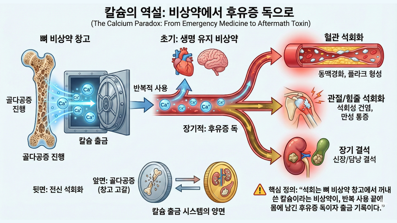
From an evolutionary perspective, what does calcium deficiency mean? In the natural environment, calcium deficiency is a signal that food is scarce. Either the environment is barren, or the individual's ability to obtain food has declined. Either way, it indicates that the individual's fitness has fallen. What response would evolution have chosen in such a situation? There are two options. First, to preserve the bone. Even when calcium is lacking, do not take it out of the bone. Blood calcium falls. The heart stops. Death follows. Second, to maintain blood calcium. Take it out of the bone. The bone weakens. But for now one can live. If food is found later, the bone can be filled again. Evolution chose the second. Immediate survival takes priority over long-term health. Because if you die today, there is no tomorrow. In the natural environment, calcium deficiency was probably usually temporary. Food availability changes with the season and the climate. If you endure the lean period, an abundant period comes. In the abundant period the bone can be filled again. But modern people's chronic calcium deficiency is different. It is not temporary but continuous. There is no recovery period. The calcium taken from the bone is not refilled. The emergency medicine becomes the everyday medicine.
Looking at the changes that come with aging, aging causes a decrease in the absorption of calcium and vitamin D, leading to deficiency; aging causes inflammaging, leading to inflammation; aging causes a decline in kidney function, leading to a tendency toward acidosis; and aging causes a decline in cardiopulmonary function, leading to a tendency toward hypoxia. Hidden within the single word aging is in fact a process in which deficiency, inflammation, acidosis, and hypoxia are all switched on gradually at the same time. Together, these four axes send the bone the signal to now draw out and use the emergency calcium.
As we saw earlier in Chapter 5, calcium absorption decreases with aging. Stomach acid secretion decreases. Among those aged 60 and over, about 10 to 30 percent are in a state of low stomach acid or achlorhydria. Stomach acid ionizes the calcium compounds in food and turns them into an absorbable form. Calcium carbonate in particular requires stomach acid to dissolve. When stomach acid is insufficient, absorption of supplements in the calcium carbonate form declines. Vitamin D synthesis in the skin decreases. In the skin, the vitamin D precursor is converted into vitamin D3 by ultraviolet B, but as one ages the precursor content in the skin declines. The skin of a 70-year-old, even when exposed to the same amount of ultraviolet light, synthesizes only about 25 percent as much vitamin D as that of a 20-year-old. The kidney's ability to activate vitamin D decreases. Vitamin D is first converted in the liver and then converted into its final active form in the kidney, but as kidney function declines with aging, this activation decreases. The intestine's capacity to transport calcium decreases. Active vitamin D increases the expression of calcium channels and calcium-binding proteins in intestinal cells, but with aging the expression of vitamin D receptors decreases and the response to the same amount of vitamin D weakens. The result of all these changes is the same. Not enough calcium comes in from the outside. The body perceives a state of deficiency. Parathyroid hormone rises. Calcium leaves the bone. Aging itself pulls the deficiency trigger.
Deficiency forms a vicious cycle. Calcium deficiency causes a rise in parathyroid hormone, the rise in parathyroid hormone causes the mobilization of calcium from the bone, the mobilization of calcium from the bone causes bone weakening, and bone weakening causes an increase in fracture risk. At the same time, vitamin D deficiency causes a decrease in calcium absorption in the intestine, the decrease in calcium absorption causes the worsening of calcium deficiency, the worsening of calcium deficiency causes a rise in parathyroid hormone, and the rise in parathyroid hormone causes accelerated bone loss. Furthermore, vitamin D deficiency causes a decline in muscle strength, the decline in muscle strength causes reduced activity, reduced activity causes a decrease in the mechanical stimulation applied to the bone, the decrease in mechanical stimulation causes reduced bone formation, and reduced bone formation causes accelerated bone loss. This is because vitamin D is also necessary for muscle function. The vicious cycles are connected to one another. Once they begin, they do not stop on their own. External intervention is needed.
2. The I Trigger: Inflammation - calcium is mobilized in the progression of inflammation
Inflammation is a double-edged sword. Acute inflammation is essential to survival. When bacteria invade, immune cells rush in, blood flow increases to produce redness and warmth, swelling forms to block the spread of pathogens, and pain arises to protect the injured area. Thanks to the inflammatory response, infection is eliminated and tissue is repaired. But when inflammation is not resolved and persists, it becomes a problem. This is chronic inflammation. Low-level inflammation persists for months, years, decades. It continuously damages tissue. Chronic inflammation is connected to almost all chronic diseases. Cardiovascular disease, diabetes, cancer, dementia, and osteoporosis. There is even a term, inflammaging, so much so that aging itself is connected to a state of chronic inflammation.
Inflammatory signaling substances directly affect bone metabolism. Tumor necrosis factor alpha is a powerful promoter of bone resorption. Tumor necrosis factor alpha activates osteoclasts through several routes. It increases RANKL expression in osteoblasts and immune cells, increases RANK expression in osteoclast precursor cells, acts directly on osteoclasts to promote their differentiation and survival, and acts on osteoblasts to suppress bone formation. Tumor necrosis factor alpha is the main cause of the loss of bone around the joints in patients with rheumatoid arthritis. Drugs that are tumor necrosis factor alpha inhibitors, such as infliximab or adalimumab, are used to treat rheumatoid arthritis, and these drugs also reduce bone loss. Interleukin-1 is also a powerful promoter of bone resorption. Interleukin-1 increases RANKL expression and decreases OPG expression. It prolongs the lifespan of osteoclasts. It also suppresses the function of osteoblasts. Interleukin-6 is one of the most commonly measured markers of inflammation. Interleukin-6 promotes osteoclast formation. The role of interleukin-6 is especially important in a state of estrogen deficiency. Estrogen suppresses the production of interleukin-6. When estrogen decreases after menopause, interleukin-6 increases, and this contributes to bone loss. Here is a point worth noting. Immune cells such as activated T cells and B cells themselves directly secrete RANKL and promote osteoclast differentiation. It is a structure in which inflammation affects bone not only through signaling substances, but in which immune cells directly flip the switch of the bone-destroying cells. The overall effect of inflammatory signaling substances is to raise the RANKL/OPG ratio. Osteoclast formation and activity increase. Bone resorption outpaces formation. Bone is lost.
Glucocorticoids, that is, steroids, which are frequently used in chronic inflammatory diseases such as rheumatoid arthritis and chronic obstructive pulmonary disease, are also harmful to bone. Steroids suppress osteoblasts and lengthen the lifespan of osteoclasts. Apart from inflammation itself, they add yet another axis of bone loss. In actual clinical practice, the combination of an inflammatory disease and steroids is one of the most powerful combinations for osteoporosis. Inflammation dissolves bone, and the drug used to treat inflammation also dissolves bone. It is a double attack.
As one ages, the baseline level of inflammatory signaling substances rises. This is called inflammaging. Several factors contribute to inflammaging. There is cellular senescence. Senescent cells stop dividing but do not die. Instead they secrete inflammatory substances. As one ages, senescent cells accumulate. These contribute to chronic inflammation. There is also increased intestinal permeability. As one ages, the intestinal barrier function weakens. Gut bacteria or bacterial products enter the bloodstream.
The immune system responds to this and provokes inflammation. There are also changes in adipose tissue. As one ages, adipose tissue increases, and the immune cells within adipose tissue increase. Adipose tissue secretes inflammatory substances such as tumor necrosis factor alpha and interleukin-6. Obesity is a state of chronic low-grade inflammation. There is also immunosenescence. As one ages, immune function declines. Paradoxically, the decline in immune function coexists with chronic inflammation. While the immune system fails to eliminate pathogens efficiently, at the same time an inappropriate inflammatory response persists. As a result of inflammaging, in the elderly, inflammatory markers such as CRP, interleukin-6, and tumor necrosis factor alpha are elevated. This elevation continuously affects the bone.
From an evolutionary perspective, what does inflammation mean? Acute inflammation is an appropriate response to infection or injury. It is essential to survival. In the natural environment, an individual with a deficient inflammatory response would have succumbed to infection. But chronic inflammation is different. In the natural environment, what became of an individual in a state of chronic inflammation? Chronic inflammation indicates that the immune system is failing to solve a problem. Either the infection persists, or the tissue damage is not healing, or the immune system is malfunctioning. Either way, the individual is not in an optimal state. An individual in a state of chronic inflammation suffers various disadvantages. Energy is consumed by the immune response. Activity declines. It becomes vulnerable to predators. Reproductive success falls. From an evolutionary perspective, chronic inflammation is a signal that there is a problem with this individual. That calcium is mobilized from the bone in response to this signal can be interpreted as a reallocation of resources from bone maintenance, a long-term investment, to the immune response and immediate survival, a short-term response. Of course, modern medicine can control chronic inflammation. Anti-inflammatory drugs, immunomodulators, biologics. But the body's basic response system remains just as it evolved. When there is an inflammatory signal, calcium leaves the bone.
The link between inflammation and bone loss is observed in several diseases. Rheumatoid arthritis is an autoimmune disease in which chronic inflammation occurs in the joints, the bone around the joints is lost, and systemic osteoporosis also occurs. The fracture risk of rheumatoid arthritis patients is about twice that of the general population. In inflammatory bowel diseases such as Crohn's disease or ulcerative colitis, chronic inflammation occurs in the intestine, the inflammation itself causes bone loss, and absorption disorders bring on calcium and vitamin D deficiency as well. The prevalence of osteoporosis among inflammatory bowel disease patients is about 15 to 40 percent. In chronic obstructive pulmonary disease there is chronic inflammation in the lungs, accompanied by systemic inflammation as well.
The prevalence of osteoporosis among chronic obstructive pulmonary disease patients is very high, about 30 to 60 percent. In chronic kidney disease, chronic inflammation accompanies the decline in kidney function, and renal osteodystrophy occurs. Diabetes is a state of chronic low-grade inflammation, and fracture risk is increased in patients with type 2 diabetes. Interestingly, fracture risk is high even though bone density may be high, because the quality of the bone declines. Periodontitis is chronic inflammation in the oral cavity, and loss of alveolar bone occurs. An association between periodontitis and systemic osteoporosis has also been reported. What these various diseases have in common is chronic inflammation. And all of them are connected to bone loss.
3. The A Trigger: Acidosis - bone is sacrificed to neutralize acid
The pH of the blood is precisely maintained at about 7.35 to 7.45. This narrow range keeps enzyme function, protein structure, cell membrane permeability, and the like in an optimal state. If the pH falls below 7.35 it is acidosis, and if the pH rises above 7.45 it is alkalosis. If the pH becomes 7.0 or lower or 7.8 or higher, life is in danger. The body regulates pH through three mechanisms. There is the chemical buffer system; the blood has several buffer systems. The most important is the carbonic acid-bicarbonate buffer system, and there are also the phosphate buffer system and the protein buffer system. These buffer systems immediately soften changes in pH. There is also respiratory regulation, in which the lungs discharge carbon dioxide. Carbon dioxide reacts with water to form carbonic acid. Breathing quickly discharges more carbon dioxide, so the pH rises; breathing slowly accumulates carbon dioxide, so the pH falls. It is regulated on the scale of minutes. There is also renal regulation, in which the kidney excretes acid and reabsorbs bicarbonate. It acts over hours to days and is the most powerful regulatory mechanism.
Here there is a fourth mechanism. It is the bone. The mineral component of bone is hydroxyapatite. This is a basic compound. It can react with acid and neutralize it. When body fluids become acidified, calcium, carbonate, and phosphate dissolve out of the bone. These basic ions neutralize the acid. The bone acts as a buffer. This happens through two routes: physicochemical dissolution and cell-mediated resorption. In physicochemical dissolution, hydroxyapatite dissolves directly in an acidic environment. It occurs chemically without the involvement of cells, and it happens relatively quickly. In cell-mediated resorption, an acidic environment activates osteoclasts. The osteoclasts resorb the bone. It is slower but more sustained. The bone is a vast alkaline reservoir. In an adult's bones there are about 1 kilogram of calcium and 0.5 kilogram of phosphorus. All of this is potential buffering capacity.
Many people receive a result at a health checkup that their blood pH is normal and feel reassured. But this may be thanks to the sacrifice of the bone. The bodies of modern people are constantly under acidifying attack from meat and processed food. The fact that the blood pH stays within the normal range of 7.35 to 7.45 may mean that the bone is desperately neutralizing the acid by shaving away its own flesh, calcium. This is called chronic latent metabolic acidosis. It is not an extreme acidosis in which the blood pH deviates greatly from the normal range, but a state in which the acid load the kidney must handle is steadily high. At this time the bone gives up its minerals little by little as it plays the role of buffer. The numbers are normal. But the bone is already dissolving. When acidosis persists, minerals continuously leave the bone. According to research, when the blood pH decreases by 0.1 unit, calcium release from the bone increases significantly. In a state of chronic metabolic acidosis, bone density decreases and fracture risk increases. Chronic kidney disease is a good example of this mechanism. When kidney function declines, the ability to excrete acid falls. Metabolic acidosis occurs. Minerals leave the bone to buffer the acid. Renal osteodystrophy occurs.
A high-protein diet also increases the acid load. When protein is metabolized, nonvolatile acids such as sulfuric acid and phosphoric acid are produced. But here there is a point of caution. A meal high in protein raises the acid load through sulfur and phosphorus metabolism, but if kidney function is normal and enough alkaline potassium is taken in together from fruits and vegetables, it is not harmful to the bone and may instead protect muscle and bone together. The problem is not protein itself so much as a dietary pattern in which fruits and vegetables are too few relative to protein. A diet of only meat and no vegetables is the problem. Conversely, an alkaline diet, that is, a diet rich in fruits and vegetables, is protective of the bone. Fruits and vegetables are rich in organic acid salts such as potassium citrate and potassium malate. When these are metabolized, they produce bicarbonate. The acid load decreases. The buffering burden on the bone is reduced.
From an evolutionary perspective, what does acidosis mean? Acidosis indicates that the metabolic balance has been broken. Either acid is produced excessively or acid excretion is insufficient. Acid is produced excessively due to metabolic abnormality or tissue hypoxia, or acid excretion is insufficient due to a decline in kidney function or a decline in lung function. Either way, there is a problem with the individual's ability to maintain homeostasis. In the natural environment, an individual in a state of acidosis would not have survived long. Acidosis is a signal of a serious internal problem. Therefore evolution chose the response of maintaining pH even at the cost of sacrificing the bone in the face of acidosis. The buffering function of the bone is advantageous for short-term survival. If the pH falls sharply, enzymes do not function. The heart can stop. Releasing minerals from the bone to maintain pH helps immediate survival. In the natural environment, acute acidosis was either resolved quickly or led to death. Surviving for a long time in a state of chronic acidosis is something that modern medicine has made possible. Through dialysis, bicarbonate supplementation, and the like, chronic kidney disease patients survive for years even in a state of acidosis. But the bone continues to be sacrificed.
The diet of modern people differs from the diet of the evolutionary environment. The diet of hunter-gatherers consisted of wild animals, wild plants, fruits, and nuts. Protein intake was high, but at the same time potassium and organic acid salt intake was also high. Wild plants and fruits are rich in potassium. The overall acid-base balance is estimated to have been neutral or mildly alkaline. The modern Western diet is different. It is high in processed food, grains, dairy, and meat. Fruits and vegetables are few. Potassium intake has decreased, and sodium and chloride intake has increased. Overall the acid load has risen. This chronic low-grade metabolic acidosis can affect bone health. The blood pH is within the normal range, but the bone continuously plays the role of buffer and loses minerals. This may be one of the reasons that eating fruits and vegetables is beneficial for bone health. Epidemiological studies show that the more fruits and vegetables one eats, the higher the bone density and the lower the fracture risk. There are also effects from calcium, vitamin K, antioxidants, and the like, but the effect of reducing the alkaline load may also contribute.