
Continuing from the previous piece, we examine the H (Hypoxia) trigger and the way the four triggers become entangled with one another. The cited references follow the manuscript as written.
4. The H Trigger: Hypoxia - When Oxygen Is Lacking, Calcium Flows Excessively into the Cell
Oxygen is the foundation of life. Every organism that carries out aerobic respiration needs oxygen. In the mitochondria, oxygen becomes the final electron acceptor of the electron transport chain. Without oxygen, the efficiency of ATP production drops sharply. From a single molecule of glucose, only 2 ATP are generated under anaerobic conditions, but with oxygen about 30 to 32 ATP are generated. When tissue lacks oxygen, energy production is impaired. Cell function declines. If it continues for a long time, the cell dies. Hypoxia arises from several causes. There are lung problems, in which the lung's gas exchange is impaired by chronic obstructive pulmonary disease, pulmonary fibrosis, pneumonia, and so on. There are heart problems, in which heart failure reduces cardiac output and so reduces oxygen delivery to the tissues. There are vascular problems, in which arteriosclerosis, thrombosis, and the like narrow or block the blood vessels so that the affected tissue becomes hypoxic. There is anemia, in which a shortage of red blood cells or hemoglobin lowers the oxygen-carrying capacity. At high altitude, low atmospheric pressure and low oxygen partial pressure produce a hypoxic state.
The cell possesses an elaborate system that senses and responds to oxygen concentration. At its center is HIF, the hypoxia-inducible factor. The 2019 Nobel Prize in Physiology or Medicine was awarded to the discoverers of this system. HIF is a transcription factor that regulates gene expression. Under normal oxygen conditions, HIF is rapidly degraded. Under hypoxic conditions, HIF is not degraded and accumulates. The accumulated HIF moves into the nucleus and regulates the expression of hundreds of genes. The genes that HIF activates help survival in a hypoxic environment. They include erythropoietin, which promotes red blood cell production; VEGF, which promotes blood vessel formation; and the enzymes that promote glycolysis.
Recent research has uncovered a startling other side of the erythropoietin that is secreted in a hypoxic state. Erythropoietin does not simply make red blood cells; it stimulates osteoclasts and dissolves bone. Why is that? The place where red blood cells are made is the bone marrow inside the bone. If oxygen is lacking and a great many red blood cells must be made in a hurry, the factory space, the bone marrow, must be expanded. So the body chooses to carve away the inner bone and secure space. When you are short of breath, bone dissolves. This is not a metaphor but a physiological fact.
Hypoxia and HIF signaling also affect bone metabolism. Research in this field is still in progress, and the results are complex. Looking at the effect on osteoclasts, osteoclasts are activated in a hypoxic environment. The bone resorption zone, the zone sealed off by the osteoclast, has a low oxygen concentration. There are studies showing that hypoxia promotes osteoclast differentiation and survival. The effect on osteoblasts is more complex. In fact, there are experiments in which animals with HIF overactivated in osteoblast-lineage cells had increased bone mass, and conversely there are studies in which bone is rapidly lost under conditions of severe systemic hypoxia or chronic disease. The effects of local, short-term hypoxia and of systemic, chronic hypoxia may differ. HIF promotes blood vessel formation through VEGF, and in bone, blood vessel formation is closely linked to bone formation. In this respect HIF may be positive for bone health. Overall, chronic hypoxia appears to have a negative effect on bone health. Clinical studies support this.
The connection between chronic hypoxic disease and bone loss is observed in several diseases. In patients with chronic obstructive pulmonary disease the prevalence of osteoporosis is very high, reported at about 30 to 60 percent. Hypoxia, chronic inflammation, glucocorticoid use, reduced activity, poor nutrition, and other factors contribute. Yet even when these factors are adjusted for, chronic obstructive pulmonary disease itself is an independent risk factor for osteoporosis. In obstructive sleep apnea, repeated hypoxia occurs, and an association between sleep apnea and osteoporosis has been reported. Intermittent hypoxia, in which breathing stops and starts again through the night as in sleep apnea, repeatedly raises sympathetic activity and oxidative stress. It is a form of hypoxic stress that goes beyond mere oxygen shortage and simultaneously stimulates hormones, inflammation, and bone metabolism. In patients with heart failure the prevalence of osteoporosis is also high. Tissue hypoperfusion and hypoxia may contribute. In chronic anemia states, especially sickle cell anemia and thalassemia, bone problems are common. Bone deformity from bone marrow hyperplasia, along with osteoporosis, occurs. Chronic hypoxia and high bone turnover contribute. The paradox of erythropoietin operates here too. There are also reports that residents at high altitude have low bone mineral density. Chronic hypoxia may contribute. However, there are other factors as well, such as physical activity and nutritional status, so the interpretation is complex.
From an evolutionary perspective, what does hypoxia mean? Hypoxia indicates that there is a serious problem with the oxygen supply. Either the lungs are not functioning properly, or the heart is not pumping properly, or the blood is not carrying oxygen. Whichever it is, there is a problem with the individual's most basic physiological function. In the natural environment, an individual in a chronic hypoxic state would not have survived long. Its capacity for activity declines. It cannot escape predators. It cannot catch prey. Its reproductive success rate falls. In response to hypoxia, HIF is activated. HIF promotes adaptations for immediate survival. It promotes glycolysis so that ATP can be produced even without oxygen. It promotes red blood cell production to raise oxygen-carrying capacity. It promotes blood vessel formation to improve oxygen delivery. These responses help short-term survival. But over the long term there is a price. Glycolysis has low efficiency and produces lactic acid. Chronic activation of HIF can promote inflammation and fibrosis. And it also affects bone metabolism. Maintaining bone in a hypoxic state may be a waste of resources. When oxygen is lacking, energy production is limited. Where should the limited energy be spent? Immediate survival, breathing, heartbeat, and brain function, take priority. Bone maintenance comes later.
The Interaction of DIAH
So far we have examined deficiency, inflammation, acidosis, and hypoxia each on their own. In reality, however, these triggers do not act independently. They are connected to one another and reinforce one another. Consider deficiency and inflammation. Deficiency of calcium and vitamin D lowers immune function. Vitamin D plays an important role in immune regulation.
Vitamin D deficiency raises susceptibility to infection and raises the risk of autoimmune disease. Infection and autoimmunity induce inflammation. Conversely, chronic inflammation lowers appetite and hinders absorption, worsening nutritional deficiency. Consider inflammation and acidosis. In an inflammatory state, metabolism changes. Glycolysis increases. Lactic acid is produced. Metabolic acidosis can be induced. Conversely, acidosis affects the inflammatory response. In an acidic environment the function of some immune cells changes.
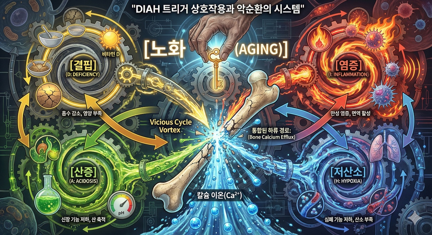
Consider acidosis and hypoxia. Tissue hypoxia induces anaerobic glycolysis. Lactic acid accumulates. Lactic acidosis occurs. Conversely, acidosis affects the oxygen dissociation curve. When pH falls, hemoglobin's oxygen affinity falls, so oxygen is supplied more readily to the tissues. This is the Bohr effect. This is a compensatory mechanism, but in a chronic state there is a complex interaction. Consider deficiency and hypoxia. Iron deficiency induces anemia. Anemia induces tissue hypoxia. Conversely, in chronic hypoxic diseases such as chronic obstructive pulmonary disease, nutritional deficiency is common due to reduced appetite and impaired absorption. In this way DIAH is connected together. When one begins, it induces or worsens the others. A vicious cycle forms. These four are like gears that turn one another, each biting the next one's tail.
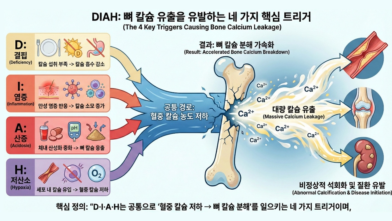
To sum up, deficiency through parathyroid hormone, inflammation through a change in the RANKL/OPG ratio, acidosis through an acidic microenvironment and direct dissolution of mineral, and hypoxia through HIF signaling and erythropoietin all converge into a single downstream pathway that leads from osteoclast activation to the efflux of calcium from bone. Four different causes produce the same result. Calcium leaves the bone. As mentioned earlier in Chapter 5, aging pulls all of the DIAH triggers. Aging causes a decrease in calcium and vitamin D absorption, which leads to deficiency; aging causes inflammaging, which leads to inflammation; aging causes a decline in kidney function, which leads to a tendency toward acidosis; and aging causes a decline in cardiopulmonary function, which leads to a tendency toward hypoxia. Hidden within the single word aging is in fact a process in which deficiency, inflammation, acidosis, and hypoxia are all switched on slowly at the same time. These four axes together send the bone the signal to take out and use the emergency calcium now.
Is this a coincidence? Or is it a design? As proposed in Chapter 5, this can be interpreted as the key of decline operating. Once reproduction ends, protective mechanisms weaken. Several systems change in the same direction at the same time. The DIAH triggers are activated. Calcium leaves the bone.
Decline proceeds. If this is a design, DIAH can be seen as a system that evaluates the state of the individual. If there is deficiency, the individual is failing to obtain food. If there is inflammation, there is damage or infection. If there is acidosis, there is a problem with metabolism. If there is hypoxia, there is a problem with breathing or circulation. If even one of these is present, the individual is not in optimal condition. If several are present, all the more so. In an aged individual all of these triggers are activated. The system judges that this individual is not in optimal condition. It withdraws resources from bone maintenance, the long-term investment.
Modern medicine can treat DIAH individually. Deficiency is treated by supplementation. Calcium supplements, vitamin D supplements, nutritional support. Inflammation is managed with anti-inflammatory treatment. Non-steroidal anti-inflammatory drugs, steroids, biologics. Acidosis is managed with bicarbonate supplementation or treatment of the cause. Sodium bicarbonate is used in chronic kidney disease. Hypoxia is treated with oxygen supply. Oxygen therapy, treatment of the underlying disease. These treatments suppress the individual triggers. But the body's basic system still operates.
When a trigger signal is present, a response that mobilizes calcium from bone takes place. This is why osteoporosis is common in chronic disease patients despite the interventions of modern medicine. An integrated approach is needed. Along with treating the individual triggers, treatment that protects the bone itself is also needed.
Conclusion
In this chapter we have examined the mechanisms of the DIAH calcium efflux triggers. Deficiency is a shortage of calcium and vitamin D. When the external supply is insufficient, parathyroid hormone mobilizes calcium from bone. The decrease in absorption that comes with aging induces deficiency. Inflammation, through inflammatory signaling molecules such as tumor necrosis factor alpha, interleukin-1, and interleukin-6, raises the RANKL/OPG ratio and activates osteoclasts. Immune cells also secrete RANKL directly. Inflammaging induces chronic inflammation. Acidosis causes the mineral of bone to be used as a buffer. Both physicochemical dissolution and cell-mediated resorption increase. A decline in kidney function and the acid load of the modern diet contribute. Even when blood pH is normal, bone may already be being sacrificed. Hypoxia affects bone metabolism through the HIF pathway. Erythropoietin stimulates osteoclasts in order to secure space inside the bone to make red blood cells. Bone loss increases in chronic lung disease, heart failure, anemia, and the like.
These DIAH triggers are not independent. They are connected to one another and form a vicious cycle. Aging activates all of the triggers at the same time. From an evolutionary perspective, DIAH can be seen as a system that evaluates the state of the individual. When the calcium efflux triggers are activated, it is a signal that the individual is not in optimal condition. In response to this signal, calcium is released from the bone. Resources are reallocated from long-term investment to short-term survival. In Chapter 7 we will examine how the effluxed calcium leads to chronic disease through the 7M mechanisms.
References
1. Harada, S., & Rodan, G. A. (2003). Control of osteoblast function and regulation of bone mass. Nature, 423(6937), 349-355. doi:10.1038/nature01660
2. Boyle, W. J., Simonet, W. S., & Lacey, D. L. (2003). Osteoclast differentiation and activation. Nature, 423(6937), 337-342. doi:10.1038/nature01658
3. Khosla, S. (2001). Minireview: the OPG/RANKL/RANK system. Endocrinology, 142(12), 5050-5055. doi:10.1210/endo.142.12.8536
4. Brown, E. M., & MacLeod, R. J. (2001). Extracellular calcium sensing and extracellular calcium signaling. Physiological Reviews, 81(1), 239-297. doi:10.1152/physrev.2001.81.1.239
5. Holick, M. F. (2007). Vitamin D deficiency. New England Journal of Medicine, 357(3), 266-281. doi:10.1056/NEJMra070553
6. Weitzmann, M. N., & Pacifici, R. (2006). Estrogen deficiency and bone loss: an inflammatory tale. Journal of Clinical Investigation, 116(5), 1186-1194. doi:10.1172/JCI28550
7. Franceschi, C., et al. (2000). Inflamm-aging: an evolutionary perspective on immunosenescence. Annals of the New York Academy of Sciences, 908(1), 244-254. doi:10.1111/j.1749-6632.2000.tb06651.x
8. Kraut, J. A., & Madias, N. E. (2010). Metabolic acidosis: pathophysiology, diagnosis and management. Nature Reviews Nephrology, 6(5), 274-285. doi:10.1038/nrneph.2010.33
9. Bushinsky, D. A. (2001). Acid-base imbalance and the skeleton. European Journal of Nutrition, 40(5), 238-244. doi:10.1007/s394-001-8352-8
10. Arnett, T. R. (2010). Acidosis, hypoxia and bone. Archives of Biochemistry and Biophysics, 503(1), 103-109. doi:10.1016/j.abb.2010.07.021
11. Semenza, G. L. (2012). Hypoxia-inducible factors in physiology and medicine. Cell, 148(3), 399-408. doi:10.1016/j.cell.2012.01.021
12. Rauner, M., et al. (2017). Perspective: hypoxia and bone. BoneKEy Reports, 6, 869. doi:10.1038/bonekey.2016.36
13. Ilich, J. Z., & Kerstetter, J. E. (2000). Nutrition in bone health revisited: a story beyond calcium. Journal of the American College of Nutrition, 19(6), 715-737. doi:10.1080/07315724.2000.10718070
14. Ginaldi, L., et al. (2005). Osteoporosis, inflammation and ageing. Immunity & Ageing, 2(1), 14. doi:10.1186/1742-4933-2-14
15. Lacey, D. L., et al. (1998). Osteoprotegerin ligand is a cytokine that regulates osteoclast differentiation and activation. Cell, 93(2), 165-176. doi:10.1016/S0092-8674(00)81569-X